All Posts By

Luciana Nofal

Cosmetic Needs Differ for Skin of Color Patients

By Media Coverage

SOURCE: Dermatology Times

If a Caucasian patient comes to my practice they will be looking for treatment for sun damage, wrinkles and capillaries, whereas, if it is a person of color, they will be looking for treatment for dark spots — dark spots from hair, dark spots from melasma, dark spots from scars, says Eliot F. Battle, Jr., M.D., CEO and Co-Founder, Cultura Dermatology and Laser Center, Washington, D.C.

Read more.

UV Protection Still Lags in Skin of Color

By Media Coverage

Source: Dermatology Times

NEW YORK ― In skin of color, ultraviolet (UV) and visible light protection are crucial to maintaining a healthy and youthful appearance. Although the mechanism by which UVA, visible light, and infrared damages the skin is still under study, reactive oxygen species (ROS) are thought to play a major role, said Steven Q. Wang, M.D., at the Skin of Color Seminar Series held here in April. Read more.

Patient received chemical peel

Staying Ahead of the Game in Skin of Color Dermatologic Care

By Sessions

The optimal treatments for skin of color patients seeking dermatologic care are constantly changing. Keeping up to date with the latest advances in the field, both medical and aesthetic, can prove to be difficult and overwhelm even the most brilliant dermatologist. With a growing recognition that constant training and direct access to skin of color thought leaders is necessary to be at the forefront of trending evidence-based research, leading experts in the field are joining forces to ensure skin of color patients receive the care they need. Among these experts are Dr. Andrew Alexis and Dr. Eliot Battle, co-chairs of the Skin of Color Seminar Series, the largest CE event dedicated to patients with skin types III – VI. With an unparalleled agenda and an esteemed faculty of KOL’s, this is probably THE event all dermatologists wanting to stay up to date on all skin of color medical and aesthetic advances must attend.

Always a highlight of the Skin of Color Seminar Series, attendees have the rare opportunity to ask their most pressing questions to the world’s top skin of color dermatology experts.

Read more.

Skin Type Classification: A Decennial Perspective

By SOC Manuscripts

Source: Journal of Drugs in Dermatology April 2018

Wendy E. Roberts MD FAAD 

The intent of this brief communication is to revisit the Roberts Skin Type Classification System published by Journal of Drugs in Dermatology (JDD) in 2008 with a 2018 lens and provide additional information for its wider acceptance and implementation. The key points of this communication are that the 2010 US census indicates rapid growth of the multiple race population up 30-50% from the 2000 census, cosmetic procedures have increased from 9.5 million to 12.8 million over the same 10 year period, and cosmetic procedures in SOC patients have increased 6% over the same 10 year period. We have come very far in our knowledge of skin safety and colorblind technology, however, as we experience rapid globalization and increasing diversity of traditionally diverse populations, this classification system is even more relevant now than it was 10 years ago. What standard are we using to predict our diverse patient outcomes to skin insult, injury, and inflammation? Why do we still use the limited Fitzpatrick Phototype System to communicate safety when the system does not address dyspigmentation and scarring, the most frequent complications in ill-fated skin trauma?

Read more.

Psoriasiform Pemphigus Foliaceus in an African American Female: An Important Clinical Manifestation

By SOC Manuscripts

Source: J Drugs Dermatol. 2018;17(4):471-473

Evan Austin BS, Jillian W. Millsop MD, Haines Ely MD, Jared Jagdeo MD MS and Joshua M. Schulman MD

A 50-year-old African-American woman presented to the dermatology clinic with a pruritic eruption of 3 years’ duration. On clinical examination, the patient had well-demarcated, pink, atrophic plaques and superficial erosions over the inframammary folds and mid-chest. She also had well-demarcated, hyperpigmented, hyperkeratotic scaly plaques over the abdomen, suprapubic region, elbows, knees, and back with sporadic small superficial blisters. A punch biopsy of the right abdomen was performed and revealed psoriasiform epidermal hyperplasia, focal parakeratosis, and acantholysis throughout the superficial spinous and granular layers. Only a sparse inflammatory infiltrate was present in the underlying dermis. Clinical and histological findings supported the diagnosis of pemphigus foliaceus (PF), but psoriasis was included in the differential diagnosis due to the presence of discrete plaques with an erythematous border. We hypothesize that patients with psoriasiform presentations of PF may be misdiagnosed with plaque psoriasis. It is important to distinguish between PF and psoriasis as there is evidence that ultraviolet light, a common treatment for psoriasis, may exacerbate PF. We document and highlight this atypical psoriasiform presentation of PF in a patient with skin of color to raise awareness and improve diagnosis and outcomes.

Read more.

alopecia lichen Planus pigmentosus

Frontal Fibrosing Alopecia and Concomitant Lichen Planus Pigmentosus: A Case Series of Seven African American Women

By Case Reports, SOC Manuscripts

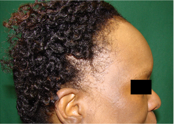
The association of frontal fibrosing alopecia (FFA) and lichen planus pigmentosus (LPPigm) is rare. Prior reports suggest that FFA and LPPigm are on the same spectrum of disease, and a diagnosis of LPPigm may predict the future development of FFA. We aim to further characterize the association between FFA and LPPigm by reviewing the clinical cases of seven African American women. Seven patients with FFA were diagnosed clinically by recession of frontotemporal hairline and confirmed by histopathologic examination showing lymphocyte-mediated cicatricial alopecia. LPPigm was diagnosed by clinical evaluation alone based on the characteristic morphology, color, and distribution of the lesions. It is difficult to distinguish whether halted progression of FFA was due to the success of the treatment regimen or spontaneous stabilization of disease over time. Our case series supports the theory that FFA and LPPigm likely exist on the same spectrum of disease. Our observations demonstrate a likely positive correlation between FFA and LPPigm.

Prior studies suggested that LPPigm may be a herald sign for FFA, predicting the future onset of frontotemporal hair loss, though there has been at least one reported case of an individual simultaneously developing FFA and LPPigm. Our results do not support the hypothesis that LPPigm is a herald sign for FFA, but do support the theory that FFA and LPPigm likely exist on the same disease spectrum. Our findings parallel that of another case series which noted that there was no obvious trend pertaining to order of FFA and LPPigm development (Figure 1). A patient with one variant of lichen planus, either FFA or LPPigm, is likely at a higher risk to develop another variant of lichen planus. FFA patients with Fitzpatrick skin type III-V may be more likely to develop LPPigm because this condition commonly presents in darker skin types. Although few studies have previously reported on FFA with concomitant LPPigm, none of these studies specifically focused on the manifestation of this phenomenon in African American women. This is particularly worth investigating, as LPPigm, when it occurs, has a strong predilection for darker skin types, and FFA is most commonly found in women.

Source: J Drugs Dermatol. 2018;17(4):397-400.

Laura N. Uwakwe MD, Leah A. Cardwell MD, Emily H. Dothard MD, Bernice I. Baroudi BS, and Amy J. McMichael MD

Read more.

Register for Skin of Color Update for more content and pearls like this article.

Skin of Color Seminar Series to Provide Latest Research and Practical Pearls for Dermatologic Treatment of Skin of Color

By Media Coverage

Source: DermWire

Now in its tenth year, the Skin of Color Seminar Series (SOCSS) will be the largest medical education event of 2018 dedicated to providing dermatologists with evidence-based research and practical pearls in treating skin of color, including patients with multiracial backgrounds. SOCSS, which will take place May 5 and 6 at the Crowne Plaza Times Square in New York, uses a didactic, case-based approach through lectures, hands-on-training, and live demonstrations. Series co-chairs and founders are dermatologists Andrew Alexis, MD and Eliot Battle, MD…

Read more.

Successful Treatment of Keloid With Fractionated Carbon Dioxide (CO2) Laser and Laser-Assisted Drug Delivery of Triamcinolone Acetonide Ointment in an African-American Man

By SOC Manuscripts

Source: J Drugs Dermatol. 2017;16(9):925-927.

Ekaterina Kraeva MD, Derek Ho MD, and Jared Jagdeo MD MS

Keloids are fibrous growths that occur as a result of abnormal response to dermal injury. Keloids are cosmetically disfiguring and may impair function, often resulting in decreased patient quality-of-life. Treatment of keloids remains challenging, and rate of recurrence is high. We present a case of a 39-year-old African-American man (Fitzpatrick VI) with a 10-year history of keloid, who was successfully treated with eight sessions of fractionated carbon dioxide (CO2) laser immediately followed by laser-assisted drug delivery (LADD) of topical triamcinolone acetonide (TAC) ointment and review the medical literature on fractionated CO2 laser treatment of keloids. To the best of our knowledge, this is the first report of successful treatment of a keloid using combination therapy of fractionated CO2 laser and LADD with topical TAC ointment in an African-American man (Fitzpatrick VI) with excellent cosmetic results sustained at 22 months post-treatment. We believe that this combination treatment modality may be safe and efficacious for keloids in skin of color (Fitzpatrick IV-VI) and other patients. This case highlights the ability of laser surgeons to safely use fractionated CO2 lasers in patients of all skin colors.

Read more.

Microneedling in All Skin Types: A Review

By SOC Manuscripts

Source: J Drugs Dermatol. 2017;16(4):308-314.

Lauren Meshkov Bonati MD, Gorana Kuka Epstein MD, and Tamara Lazic Strugar MD

Microneedling procedures are growing in popularity for a wide variety of skin conditions. This paper comprehensively reviews the medical literature regarding skin needling efficacy and safety in all skin types and in multiple dermatologic conditions. A PubMed literature search was conducted in all languages without restriction and bibliographies of relevant articles reviewed. Search terms included: “microneedling,” “percutaneous collagen induction,” “needling,” “skin needling,” and “dermaroller.” Microneedling is most commonly used for acne scars and cosmetic rejuvenation, however, treatment benefit has also been seen in varicella scars, burn scars, keloids, acne, alopecia, and periorbital melanosis, and has improved flap and graft survival, and enhanced transdermal delivery of topical products. Side effects were mild and self-limited, with few reports of post-inflammatory hyperpigmentation, and isolated reports of tram tracking, facial allergic granuloma, and systemic hypersensitivity.

Read more.