All Posts By

Dana Turner

Thank You to Pfizer for Supporting Skin of Color Update 2021

By Uncategorized

Please take a moment to acknowledge and thank our gracious supporter, Pfizer, by visiting their sponsor page on the Skin of Color Update 2021 Virtual Platform and their website below

Pfizer Inc.: Breakthroughs that change patients’ lives
At Pfizer, we apply science and our global resources to bring therapies to people that extend and significantly improve their lives. We strive to set the standard for quality, safety and value in the discovery, development and manufacture of health care products, including innovative medicines and vaccines. Every day, Pfizer colleagues work across developed and emerging markets to advance wellness, prevention, treatments and cures that challenge the most feared diseases of our time. Consistent with our responsibility as one of the world’s premier innovative biopharmaceutical companies, we collaborate with health care providers, governments and local communities to support and expand access to reliable, affordable health care around the world. For more than 150 years, we have worked to make a difference for all who rely on us. We routinely post information that may be important to investors on our website at www.pfizer.com. In addition, to learn more, please visit us on www.pfizer.com and follow us on Twitter at @Pfizer and @Pfizer_News, LinkedIn, YouTube and like us on Facebook at Facebook.com/Pfizer.

Thank You to Abbvie for Supporting Skin of Color Update 2021

By Uncategorized

Please take a moment to acknowledge and thank our gracious supporter, Abbvie, by visiting their sponsor page on the Skin of Color Update 2021 Virtual Platform and their website below.

Abbvie Logo

AbbVie’s mission is to discover and deliver innovative medicines that solve serious health issues today and address the medical challenges of tomorrow. We strive to have a remarkable impact on people’s lives across several key therapeutic areas: immunology, oncology, neuroscience, eye care, virology, women’s health and gastroenterology, in addition to products and services across our Allergan Aesthetics portfolio.

Thank You to Unilever for Supporting Skin of Color Update 2021

By Uncategorized
Please take a moment to acknowledge and thank our gracious supporter, Unilever, by visiting their sponsor page on the Skin of Color Update 2021 Virtual Platform and their website below
At Unilever we believe in Positive Beauty. A vision of beauty that’s equitable, inclusive and sustainable, with brands that not only do less harm, but also do more good for people and for the planet with brands such as Dove, Mele and Vaseline. Through our beauty and personal care brands, we’re taking action to drive positive change. We’re setting out to transform the systems that hold individuals back – by advocating new policies, laws and social norms that will promote inclusion, health and wellbeing for all members of society. We’re embracing a new era of beauty. One that’s inclusive. Equitable. Regenerative. Positive. So that people and the planet can thrive together.
At Dove, we have a vision of a world where beauty is a source of confidence, not anxiety. Our mission is to ensure the next generation grow up enjoying a positive relationship with the way they look.  Dove is committed to helping women realize their personal potential for beauty by engaging them with products that deliver real care. From self-esteem and body image to ending discrimination against black hair, Dove is working to make positive changes for our people and planet.
Introducing MELĒ, a first of its kind national skincare brand that is specifically designed for, and co-created in collaboration with, women and experts with melanin-rich skin. We are a science-led brand developed with and for people with melanin-rich skin with one purpose in mind – ensure melanin-rich skin is treated as a priority and never an afterthought. With the help of dermatologists of color and a community lab with influencers and skincare enthusiasts from the community, we co-developed a range of products that focus on the most common skin needs and concerns of melanin-rich skin.
For over 150 years, Vaseline has been committed to helping heal skin everywhere and we are on a mission to ensure people of color get the quality care they deserve. We believe all people should have equal access to care – for their wellbeing and their lives. In partnership with Hued, Vaseline® introduces a new Dermatologist finder tool that allows people of color to identify and connect with dermatologists of color and those experienced in treating skin of color.

Thank You to Scientis for Supporting Skin of Color Update 2021

By Uncategorized

Please take a moment to acknowledge and thank our gracious supporter, Scientis, by visiting their sponsor page on the Skin of Color Update 2021 Virtual Platform and their website below.

Scientis is an emerging Swiss dermatology company dedicated to discovering, developing and bringing novel dermo-cosmetic products to market for skin pigmentation disorders.
With Cyspera®, the first and only topical cream with cysteamine and free of hydroquinone, we offer uncompromising scientific efficacy coupled with long-term safety benefits.

Skin of Color Update Expands in 2021

By Uncategorized
New York (June 17, 2021) – Skin of Color Update, the largest medical education event focused on the dermatologic treatment of patients with skin of color, is expanding in 2021 with an additional half day of programming and pre-conference symposium. The event will be held virtually September 10-12 with the symposium on August 3.

Read More

SANOVAWORKS TO MATCH CMMP GRANTS FOR MEDICAL STUDENTS

By Uncategorized
CMMP Medical School Grant

SanovaWorks has developed a partnership with and in support of Comprehensive Medical Mentoring Program. We are happy to announce that SanovaWorks will match the CMMP Medical Student Grant fund, which was started to help medical students from underrepresented populations with residency preparation expenses. This matching donation will ensure that more students are able to benefit from the grant. We join other partners in support of CMMP’s efforts of mentoring students, promoting diversity in medicine, and giving back to the communities.

READ MORE ABOUT CMMP’S PARTNERSHIPS


Medical Students, Grants are Now Available for Residency Expenses
Comprehensive Medical Mentoring Program (CMMP) is aware that applying to residency can be a daunting process which can also become more stressful given the associated expenses that come with it.

CMMP is providing grants up to $1,000 for rising 4th-year minority medical students as they are preparing for away rotations and residency applications.

IF YOU ARE

    1. a rising 4th-year medical school student, and
    2. applying for residency for the 2022-2023 academic year, and
    3. a member of an underrepresented group in medicine,

SUBMIT your application today!


The application deadline is May 15, 2021


Apply Today:
 https://sanovaworks.com/CMMP-medical-student-grant


JOIN US
SanovaWorks joins institutions that include George Washington University School of Medicine and Health Sciences, Howard University Hospital, Department of Dermatology, Georgetown University School of Medicine, and many others in support of CMMP.

Do you or an organization you know want to add your support to the Medical Student Grant Fund?

Contact Dr. Lauren Payne to add your company’s name to the partnership list: https://www.cmmpmed.org/contact-us/ 

Director of Equity, Diversity and Inclusion (DEDI) Call for Applications

By Uncategorized
Director of Equity, Diversity and Inclusion (DEDI) Call for Applications
ODAC Dermatology, Aesthetic & Surgical Conference has announced a call for applications for the role of Director of Equity, Diversity and Inclusion (DEDI). This newly defined position will work closely with the ODAC Advisory Board and the @SanovaWorks_ Events Division, to promote the development and implementation of educational offerings aimed at improving cultural competence, diversity, and equity within the field of dermatology. Additionally, this role will assist in creating a conference experience conducive to a collaborative learning environment that enhances professional development and advancement for all.
To qualify for consideration, applicants should possess the following experience and skills:
·    Board-certified dermatologist, 3-5 years out of residency preferred
·    Demonstrated knowledge of inclusive dermatological education that is representative of diverse patient populations
·    Training or teaching facilitation in the field of dermatology
·    Demonstrated leadership role experience with the ability to work independently
·    Commitment to promoting diversity in dermatology as evidenced by current work in healthcare community engagement
·    Strong collaborative skills with the ability to facilitate dialogue and engage in difficult conversations to sustain a culturally responsive environment
·    Excellent written and oral communication skills with knowledge of language sensitive to inclusivity of all backgrounds and communities
·    The Director of Equity, Diversity and Inclusion may not concurrently serve in the same role/capacity at other dermatology conferences

SKIN OF COLOR UPDATE PRE-CONFERENCE VIRTUAL SYMPOSIUM

By Medical Dermatology, Sessions, Skin of Color Update Agenda
A Case-Based Conversation with The Experts: Treating Pigmentary Disorders in Skin of Color Patients
SKIN OF COLOR UPDATE PRE-CONFERNECE VIRTUAL SYMPOSIUM
TUESDAY, AUGUST 3RD | 6:00PM ET – 9:00PM ET
The Skin of Color Update invites you to join its pre-conference symposium where co-chairs Drs. Andrew Alexis and Eliot Battle will host an interactive, case-based conversation with pigmentary disorders experts. Faculty will review treatment options for common as well as challenging and less frequently discussed pigmentary conditions in skin of color patients. Through a detailed review of each case, panelists will provide guidance and evidence-based treatment protocols as well as practical pearls drawn from their clinical experience. You will walk away from this session armed with clinical pearls immediately useful in your practice. In addition, all panelists will participate in live Q&A sessions to answer your most pressing questions about treating pigmentary disorders in SOC.
AGENDA
6:00-6:05 PM – Welcome & Introductions from Symposium Moderators – Andrew F. Alexis, MD, MPH & Elliot F. Battle, MD
6:05-6:20 PM – A Challenging Case of Melasma – Heather Woolery-Lloyd, MD
6:20-6:35 PM – A Case of Vitiligo Treated with Pulsed Corticosteroids/JAK-Inhibitor – Seemal Desai, MD
6:35-6:50 PM – Post-Inflammatory Hyperpigmentation(PIH) Topical & Procedural Treatment – Neelam Vashi, MD
6:50-7:00 PM – Live Audience Q&A
7:00-8:00PM – For Patients with Plaque Psoriasis: An Oral, Non-Biologic Therapy With Data on Clearer Skin and Symptoms – Paul Wallace, MD, MPA (Non-CE Workshop)
8:00-8:05 PM – Welcome & Introductions from Symposium Moderators – Andrew F. Alexis, MD, MPH & Elliot F. Battle, MD
8:05-8:20 PM – A Challenging Case of Erythema Dyschromicum Perstans – Nada Elbuluk, MD, MSc
8:20-8:35 PM – A Case of Hypopigmented Mycosis Fungoides – Eva Kerby, MD
8:35-8:50 PM – Lichen Planus Pigmentosus – Mukta Sackdev, MD
8:50-9:00 PM – Live Audience Q&A
SYMPOSIUM CO-CHARIS
Andrew F. Alexis, MD, MPH
Eliot F. Battle, MD
EXPERT FACULTY
Seemal R. Desai, MD, FAAD
Nada Elbuluk MD, MSc
Eva Kerby, MD
Heather Woolery-Lloyd, MD
Mukta Sachdev, MD
Neelam Vashi, MD

Treating Alopecia in Skin of Color Patients: How to Arrest Loss and Promote Regrowth

By Uncategorized
Treating Alopecia in Skin of Color Patients

Source: Next Steps in Derm

Can you improve your culturally-competent hair loss consultation? Would you like to grow your toolbox for comprehensive alopecia treatment? If so, you are in the right place!

Hair loss is a frequent concern for patients visiting the dermatologist, especially in patients with pigmented skin types. Kinky or coiled hair has an innate fragility that makes African Americans especially prone to hair loss concerns. At the  2020 Skin of Color Virtual Update, Dr. Susan Taylor discussed Conventional Treatment Approaches for Hair & Scalp Disorders in Skin of Color Patients. Dr. Taylor advised how to have a culturally competent hair loss consultation, reviewed common patterns of hair loss, and highlighted comprehensive treatment of multifactorial causes of alopecia.

Before we get into therapeutic strategies for alopecia, let’s highlight important considerations to institute an effective treatment plan:

  • Understand common ethnic hairstyles and practices
  • Recognize the impact of hair and scalp disorders
  • Clearly identify the problem and obtain a thorough history, including details regarding hair care practices, products used, and symptoms
  • Complete physical examination and consider scalp biopsy
  • Accurately diagnose the disease (see Table 1 for common causes of alopecia)
  • Effectively and competently partner with the patient to treat the disorder with appropriate therapy
Common Cause of Scarring Non-Scarring Alopecia

Dr. Taylor stressed the importance of culturally competent questioning during the visit. For example, asking an African American woman if she shampoos daily can reduce the patient’s confidence in the physician’s understanding of her hair type, especially during a race discordant visit. To build confidence, it is critical to understand routine hair practices and to be familiar with common products.

During the hair loss consultation, it is important to understand the problem experienced by the patient. Frequent symptoms noted by the patient that can clue in the underlying alopecia include:

    • Hair length is not increasing.
    • There is hair breakage.
    • Hair is falling out at the root.
    • There is decreased hair density.
    • There is focal or localized hair loss.

Other considerations include the duration of hair loss, any associated symptoms, and presence of family history of hair loss. Further, it is important to ask about their hair care history. The types of hairstyles used by the patient and the hair care routine should be discussed.

The physical exam should be thorough to evaluate the scalp and hair density, a hair pull test, and examination of the eyebrows and eyelashes. The presence of weaves, braids, or wig caps may impede evaluation. If the patient has any of these hairpieces on the scalp that cannot be easily removed during the visit, the patient should return for evaluation after these are removed. History can still be discussed during the visit, however, the co-pay should be refunded, and the patient should be offered an earlier appointment for evaluation of the hair and scalp.

Now that we have discussed how to approach the consultation, let’s get into the alopecias! Dr. Taylor highlighted central centrifugal cicatricial alopecia, traction alopecia, and frontal fibrosing alopecia, as well as tips to minimize breakage, reduce hair tension, and scalp care to treat seborrheic dermatitis.

Central centrifugal cicatricial alopecia (CCCA) is a scarring alopecia seen in about 5.6% of African American women. It frequently starts with increased hair breakage at the vertex, and patients often have associated symptoms of itching, burning, tenderness, and soreness of the scalp. The goals of treatment are to aggressively treat the CCCA in the early stages to prevent progression, relieve symptoms, and possibly have some regrowth in the affected areas. There are no randomized controlled trials for CCCA treatment, thus evidence for treatment comes from case reports and series. The mainstay of treatment is corticosteroids (topical and intralesional), anti-inflammatory antibiotics, antimalarials, and minoxidil (topical and oral formulations). Anti-seborrheic shampoos, hair transplantation, and platelet-rich plasma injections serve as adjuncts to treatment. See Table 2 for detailed treatment options. Frequently multiple agents are used in combination to control the disease.

Two important areas of focus when treating CCCA that Dr. Taylor highlighted are practices to minimize hair breakage and treatment of frequently concurrent seborrheic dermatitis.

Read More…..

Cutaneous Sarcoidosis

Hypopigmented Cutaneous Sarcoidosis Responsive to Minocycline

By Case Reports, SOC Manuscripts

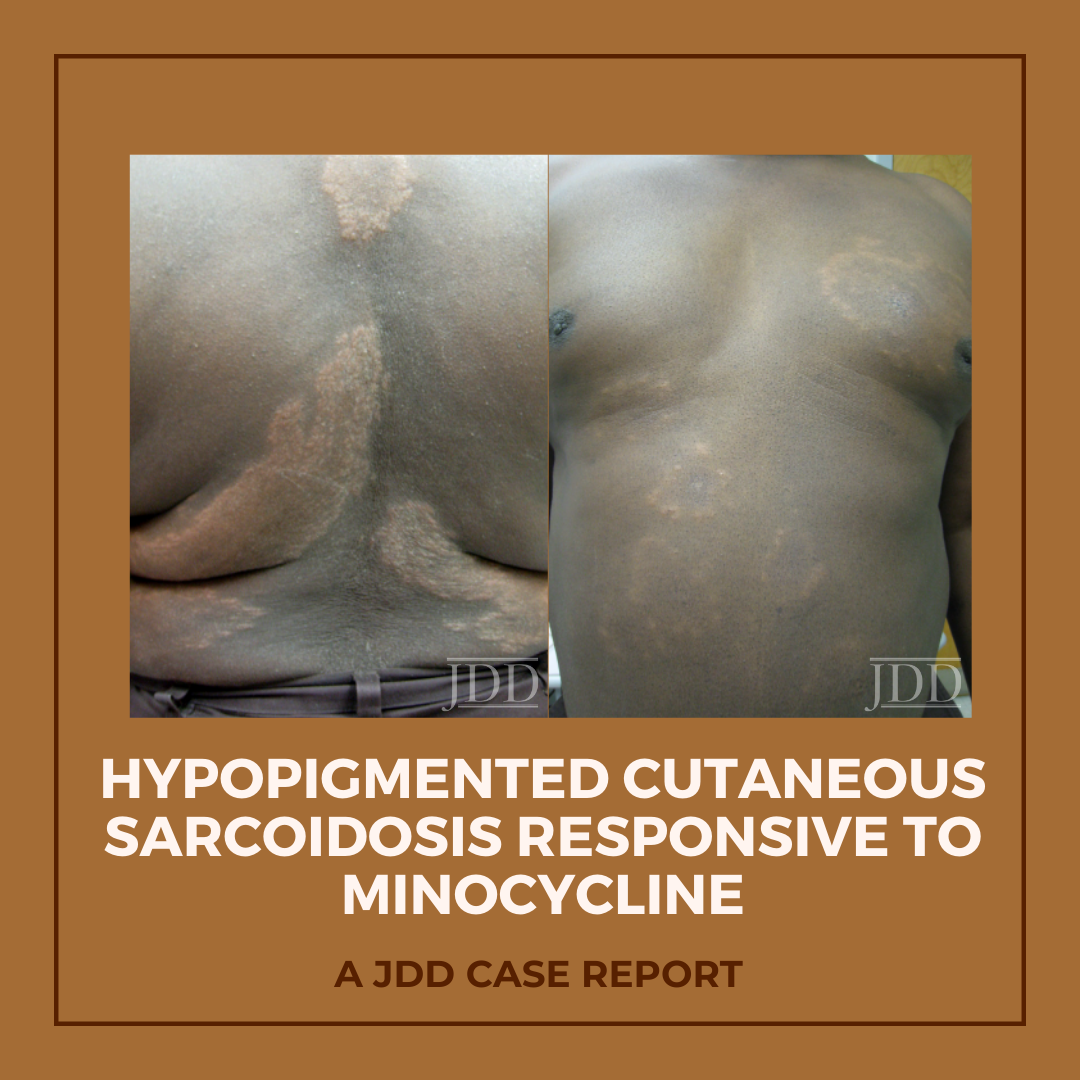
Hypopigmented patches and plaques are a rare presentation of cutaneous sarcoidosis. JDD authors describe a case of generalized hypopigmented cutaneous sarcoidosis that showed good response to minocycline therapy.

Introduction

A 58-year-old African-American male with a past medical history of hypertension, diabetes mellitus, tobacco use, glucose-6-phosphate dehydrogenase (G6PD) deficiency, and hyperlipidemia presented with a two-year history of asymptomatic light spots on his trunk and upper extremities. He reported a history of cutaneous sarcoidosis a decade prior, characterized by erythematous papules and plaques that had regressed with hydroxychloroquine therapy. The newer light patches were not responsive to mid-potency topical steroids or tacrolimus 0.1% ointment. Review of systems was negative. The patient denied any new medications or history of travel outside of the metropolitan area.


Physical examination of the skin was significant for multiple hypopigmented patches on the face, neck, and extremities; hypopigmented plaques on the back (Figure 1); and annular plaques of hypopigmented papules on the chest and abdomen (Figure 2). The lesions were not hypoesthetic. There was no lymphadenopathy and physical exam was otherwise unremarkable.


A biopsy from the left arm showed a superficial and deep multinodular granulomatous infiltrate sparing the epidermis (Figure 3). The granulomas were predominantly composed of epithelioid histiocytes with a few scattered lymphocytes (Figure 4). Special stains for microorganisms were negative. There was no appreciable epidermal change or pigment incontinence. The histopathological picture was consistent with recurrent cutaneous sarcoidosis.
Computed tomography of the chest with and without contrast showed no evidence of active pulmonary sarcoidosis or lymphadenopathy. An ophthalmologic exam, abdominal ultrasound, and spirometry were within normal limits, as were serum cal cium and angiotensin-converting enzyme (ACE) levels.


Because the patient’s skin manifestations had resolved with hydroxychloroquine in the past, this treatment was restarted at 200 mg twice-daily. Unfortunately, laboratory monitoring revealed hepatic transaminitis and mild anemia 2 months into the treatment course, corresponding with only minimal improvement of the hypopigmented plaques, necessitating discontinuation of hydroxychloroquine. Minocycline at a dose of 100 mg twice daily was then initiated 4 months after normalization of liver function tests. After 5 months of treatment, all hypopigmented patches, papules, and plaques had completely or partially repigmented and were appreciably smoother and flatter (Figures 5 and 6). The patient tolerated the medication well with no adverse effects.


Cutaneous Sarcoidosis

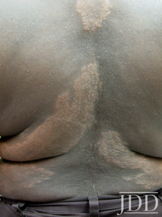
FIGURE 1. Hypopigmented plaques over the back.

Cutaneous Sarcoidosis

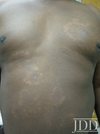
FIGURE 2. Hypopigmented annular plaques of papules on the abdomen.

Cutaneous Sarcoidosis

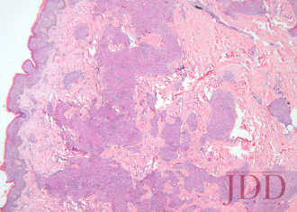
FIGURE 3. Hematoxylin and eosin stain of a left arm skin biopsy show-ing dermal granulomatous inflammation.

Cutaneous Sarcoidosis

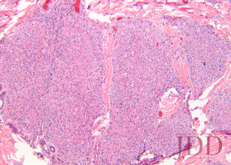
FIGURE 4. Hematoxylin and eosin stain of a left arm skin biopsy at higher magnification demonstrating well-formed naked granulomas.

Cutaneous Sarcoidosis

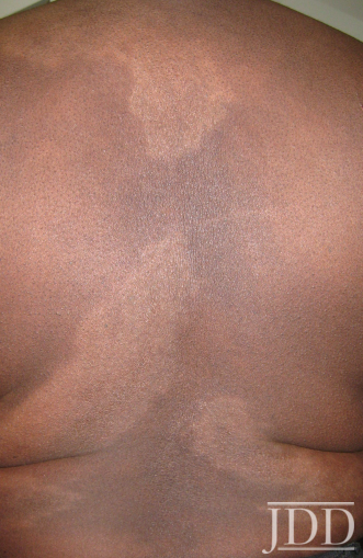
FIGURE 5. Flattening and repigmentation of plaques on the back after treatment with minocycline.

Cutaneous Sarcoidosis

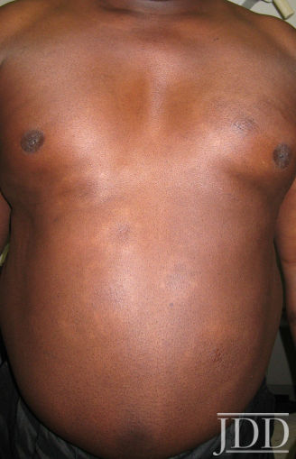
FIGURE 6. Repigmentation of annuli on the anterior trunk after treat-ment with minocycline.


Discussion

The skin is one of myriad organs potentially affected by sarcoidosis, a multisystem idiopathic disorder characterized histologically by infiltration of noncaseating granulomas. Cutaneous manifestations of sarcoidosis are protean, including papules and plaques of various morphology and distribution, subcutaneous nodules, pruritus, ichthyosis, erythroderma, ulceration, verrucosis, nail disease, and infiltrative scars.1 In the United States, sarcoidosis is more common in African-Americans than in other ethnic groups, and cutaneous manifestations in individuals of African descent are more likely to be atypical.2

In 1973, Cornelius et al reported 4 patients who presented with hypopigmented and depigmented patches and plaques that showed the naked tuberculoid granulomas characteristic of sarcoidosis on skin biopsy.3 The distribution in these cases was variable, with generalization in one patient and localization to the face, legs only, or legs and arms in the other three. Histologically, no difference in epidermal melanocyte count was noted between affected and unaffected skin, but relative hypomelanosis of the malphigian and corneal layers was appreciated. All patients had multisystem disease. Four years later, Hubler described a “hypomelanotic canopy” in a woman with lupus pernio and lymphadenopathy whose hypopigmented patches overlay deep subcutaneous nodules on the arms.4Biopsy of an enlarged lymph node revealed epithelioid granulomas that were reduplicated on skin biopsy. Interestingly, cutaneous hypopigmentation lacking any histological evidence of granulomatous dermatitis has also been observed in the setting of systemic sarcoidosis;5,6 this highlights the need for vigilant continued surveillance when the clinical index of suspicion for sarcoidosis is high and biopsy is noncorroborative.


The etiopathogenesis of hypopigmentation in sarcoidosis, like the disease itself, is unknown. Theories regarding the likely mul tifactorial cause of sarcoidosis center around T-cell—mediated autoimmunity, genetic predisposition, and aberrant response to bacterial or other antigens; the evidence for each has been extensively reviewed elsewhere.1,7 The rare cutaneous hypopigmentation seen in dark-skinned individuals with sarcoidosis appears to represent a melanopenic hypomelanosis, as opposed to postinflammatory hypopigmentation or melanocytopenia.3,4,8 In our patient, there were no epidermal changes, interface in flammation, or melanin-containing dermal macrophages to suggest postinflammatory dyspigmentation.


Minocycline represents an attractive alternative treatment for chronic sarcoidosis in patients who might otherwise be treated with long-term corticosteroids, antimalarials, methotrexate, or other immunosuppressants. The tetracycline class of antibiotics has previously demonstrated utility in a variety of dermatoses and autoimmune-connective tissue diseases, and may be particularly efficacious for granulomatous skin conditions.9,10 Tetracyclines have specifically shown efficacy in the treatment of granulomatous periorificial dermatitis, cheilitis granulomatosa, granulomatous rosacea, and silicone granulomas.11-14 More recently, tetracyclines proved beneficial in treating granuloma annulare (GA), an entity often with significant clinicopathological overlap with cutaneous sarcoidosis.15,16 Marcus et al showed that, in combination with rifampin and ofloxacin, 3 to 5 courses of monthly minocycline therapy led to complete clearance of GA in 6 patients, half of whom had generalized disease.15 In this series, the rationale for selection of the same triple combination antimicrobial therapy as that used for paucibacillary leprosy (PBL) was that the clinical and histological similarities between PBL and GA might impart an equally similar response to treatment. The same logic could be extrapolated further to include patients with cutaneous sarcoidosis as candidates for a treatment regimen, including minocycline, with or without rifampin and ofloxacin.


Minocycline as monotherapy was efficacious for the treatment of chronic cutaneous sarcoidosis in an open observational study of 12 patients by Bachelez et al.17 The cutaneous manifestations of patients in the Bachelez group included classic plaques, papulonodules, subcutaneous nodules, and lupus pernio. Ten of 12 patients completely or partially responded to treatment, which was generally well tolerated except for drug hypersensitivity syndrome in one patient with a history of other autoimmune diseases. Park et al later reported a patient with cutaneous, lacrimal gland, pulmonary, and ocular (choroidal) sarcoidosis that responded to minocycline.18

Miyazaki et al described a patient with a unique presentation of muscular sarcoidosis of the limbs, as well as uveitis and pulmonary disease, in whom clinical response to minocycline was paralleled by normalization of an elevated ACE level.19 The patient relapsed off of minocycline, but rapidly responded to reintroduction of the medication. Immunohistochemical staining of muscle biopsy specimens for Propionibacterium acnes showed multiple small particles within granuloma macrophages and giant cells, substantiating the theory that sarcoidosis is an infectious disease or an aberrant immunologic response to bacteria. Evidence for the role of other infectious agents in sarcoidosis, especially Mycobacterium tuberculosis, has been mixed and inconclusive.20 That sarcoidosis is purely an infectious disease is certainly within the realm of possibility,21 but the recurrence of disease once antibiotics are discontinued argues against this theory.19 An alternative explanation is that the benefits of tetracyclines in sarcoidosis and other autoimmune diseases are derived more from their nonantibiotic immunomodulating properties. The anti-inflammatory properties ascribed to minocycline include inhibition of T cell activation, proliferation, and transmigration as well as expression of nitric oxide synthetase and matrix metallopeptidase 9 (MMP-9).9Furthermore, tetracyclines have been shown to inhibit granuloma formation in vitro.22


Conclusion

The role of tetracyclines in the therapeutic armamentarium of sarcoidosis, especially in those who cannot tolerate antimalarials or other immunomodulating medications, is likely to expand. Whether minocycline is particularly effective for the hypopigmented variety of cutaneous sarcoidosis remains to be seen.


Disclosures

The authors have no relevant conflicts of interest to disclose.

References

  1. English JC 3rd, Callen JP. Sarcoidosis. In: Callen JP, Jorizzo JL, Bolognia JL, et al, eds. Dermatological Signs of Internal Disease. 4th ed. China: Elsevier; 2009:287-295.
  2. Jayck WK. Cutaneous sarcoidosis in black South Africans.Int J Dermatol. 1999;38(11):841-845.
  3. Cornelius CE 3rd, Stein KM, Hanshaw WJ, Spott DA. Hypopigmentation and sarcoidosis. Arch Dermatol.1973;108(2):249-251.
  4. Hubler WR Jr. Hypomelanotic canopy of sarcoidosis. Cutis.1977;19(1):86-88.
  5. Alexis JB. Sarcoidosis presenting as cutaneous hypopigmentation with repeatedly negative skin biopsies.Int J Dermatol. 1994;33(1):44-45.
  6. Hall RS, Floro JF, and King LE Jr. Hypopigmented lesions in sarcoidosis. J Am Acad Dermatol. 1984;11(6):1163-1164.
  7. English JC 3rd, Patel PJ, Greer KE. Sarcoidosis. J Am Acad Dermatol. 2001;44(5):725-743.
  8. Clayton R, Breathnach A, Martin B, et al. Hypopigmented sarcoidosis in the Negro. Report of eight cases with ultrastructural observations. Br J Dermatol.1977;96(2):119-125.
  9. Sapadin AN, Fleischmajer R. Tetracyclines: nonantibiotic properties and their clinical implications. J Am Acad Dermatol. 2006;54(2):258-265.
  10. Stone M, Fortin PR, Pacheco-Tena C, Inman, RD. Should tetracycline treatment be used more extensively for rheumatoid arthritis? Metaanalysis demonstrates clinical benefit with reduction in disease activity J Rheumatol.2003;30(10):2112-2122.
  11. Falk ES. Sarcoid-like granulomatous periocular dermatitis treated with tetracycline. Acta Derm Venereol.1985;65(3):270-272.
  12. Camacho F, García-Bravo B, and Carrizosa A. Treatment of Miescher’s cheilitis granulomatosa in Melkersson-Rosenthal syndrome. J Eur Acad Dermatol Venereol.2001;15(6):546-549.
  13. Mullanax MG, Kierland RR. Granulomatous rosacea. Arch Dermatol. 1970;101(2):206-211.
  14. Senet P, Bachelez H, Ollivaud L, Vignon-Pennamen D, Dubertret L. Mi- nocycline for the treatment of cutaneous silicone granulomas. Br J Dermatol. 1999;140(5):985-987.
  15. Marcus DV, Mahmoud BH, Hamzavi IH. Granuloma annulare treat- ed with rifampin, ofloxacin, and minocycline combination therapy Arch Dermatol. 2009;145(7):787-789.
  16. Duarte AF, Mota A, Pereira MA, Baudrier T, Azevedo F. Generalized granuloma annulare—response to doxycycline.J Eur Acad Derma- tol Venereol. 2009;23(1):84-85.
  17. Bachelez H, Senet P, Cadranel J, Kaoukhov A, Dubertret L. The use of tetracyclines for the treatment of sarcoidosis.Arch Dermatol. 2001;137(1):69-73.
  18. Park DJ, Woog JJ, Pulido JS, Cameron JD. Minocycline for the treatment of ocular and ocular adnexal sarcoidosis.Arch Ophthal- mol. 2007;125(5):705-709.
  19. Miyazaki E, Ando M, Fukami T, Nureki S, Eishi Y, Kumamoto T. Mi- nocycline for the treatment of sarcoidosis: Is the mechanism of action immunomodulating or antimicrobial effect? Clin Rheumatol. 2008;27(9):1195-1197.
  20. Tchernev G. Cutaneous sarcoidosis: the “great imitator”: etiopathogenesis, morphology, differential diagnosis, and clinical management. Am J Clin Dermatol. 2006;7(6):375-382.
  21. Marshall TG, Marshall FE. Sarcoidosis succumbs to antibiotics—implications for autoimmune disease.Autoimmun Rev.
  22. Webster GF, Toso SM, and Hegemann L. Inhibition of a model of in vitro granuloma formation by tetracyclines and ciprofloxacin. Involvement of protein kinase C. Arch Dermatol. 1994;130(6):748-752.

Originally published in the Journal of Drugs in Dermatology in April 2012. 

Schmitt, C. E., Fabi, S. G., Kukreja, T., & Feinberg, J. S. (2012). Hypopigmented cutaneous sarcoidosis responsive to minocycline. Journal of drugs in dermatology: JDD, 11(3), 385-389. https://jddonline.com/articles/dermatology/S1545961612P0385X  

Content and images republished with permission from the Journal of Drugs in Dermatology.

Adapted from original article for length and style.

The Journal of Drugs in Dermatology is available complimentary to US dermatologists, US dermatology residents, and US dermatology NP/PA. Create an account on JDDonline.com and access over 15 years of PubMed/MEDLINE archived content.

Did you enjoy this case report? You can find more here.