All Posts By

Dana Turner

Pemphigus Foliaceus

Psoriasiform Pemphigus Foliaceus in an African American Female: An Important Clinical Manifestation

By Case Reports, SOC Manuscripts

JDD authors document and highlight this atypical psoriasiform presentation of Pemphigus Foliaceus in a patient with skin of color to raise awareness and improve diagnosis and outcomes.

Case Report

A 50-year-old African-American woman presented to the dermatology clinic with a pruritic eruption of 3 years’ duration that began as discrete plaques on the inframammary folds and subsequently spread towards the mid-chest, ears, back, elbows, knees, and scalp. Past treatments by other clinicians included clotrimazole cream and a topical corticosteroid of unknown potency without significant improvement. She denied any new medications and was taking aspirin, divalproex, mirtazapine, cetirizine, venlafaxine, atorvastatin, and omeprazole.

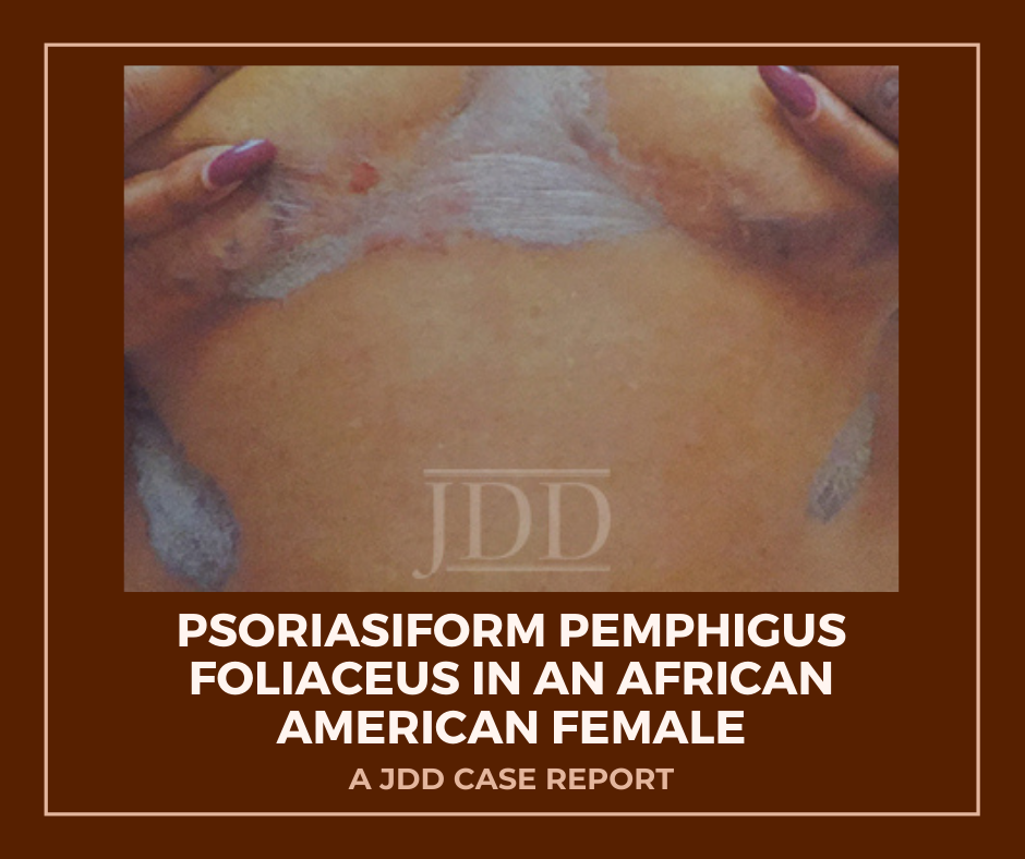
On clinical examination, the patient had well-demarcated, pink, atrophic plaques and superficial erosions over the inframammary folds and mid-chest (Figures 1). She also had well-demarcated, hyperpigmented, hyperkeratotic scaly plaques over the abdomen, suprapubic region, elbows, knees, and back with sporadic small superficial blisters (Figure 2). Complete blood count, complete metabolic panel, rheumatoid factor, and antinuclear antibody were within normal limits. Rapid plasma reagin test was negative. Erythrocyte sedimentation rate was elevated at 54 millimeter/hour (reference range 0-22 millimeter/hour). A punch biopsy of the right abdomen was performed and revealed psoriasiform epidermal hyperplasia, focal parakeratosis, and acantholysis throughout the superficial spinous and granular layers (Figure 3). Only a sparse inflammatory infiltrate was present in the underlying dermis. These clinical and histological findings supported the diagnosis of pemphigus foliaceus (PF). Patient was started on 50 mg oral dapsone daily.

Pemphigus Foliaceus

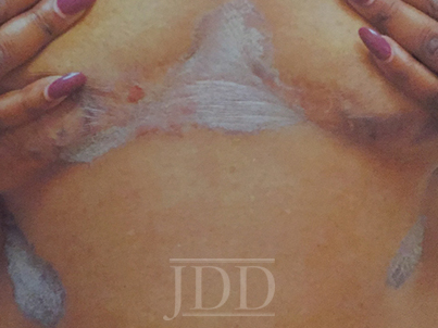
FIGURE 1. Well-demarcated, scaly plaques over the mid-chest and inframammary folds, with a single superficial erosion on the right medial chest.

Pemphigus Foliaceus

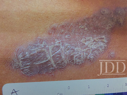
FIGURE 2. A plaque from the abdomen demonstrates “corn flake-like” scale.

Pemphigus Foliaceus

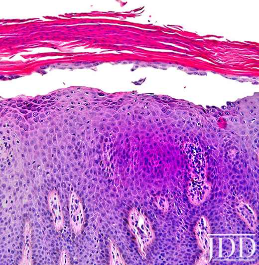
FIGURE 3. Composite photomicrograph, hematoxylin, and eosin, original magnification x200.


Discussion

Herein, we present a case of Pemphigus Foliaceus with a psoriasiform clinical presentation in an African-American patient. PF is an autoimmune skin disease caused by antibodies against the desmosomal glycoprotein, desmoglein 1.¹ Desmogleins, members of the cadherin family, serve to anchor epidermal desmosomes between adjacent keratinocytes and assist in epithelial differentiation.² Antibodies targeting desmoglein 1 result in acantholysis in the upper epidermis with limited separation in the basal layers and minimal mucosal involvement as desmoglein 1 is primarily expressed in the granular layer of the non-mucosal epidermis.¹ Patients present with scaly plaques on an erythematous base and fragile shallow blisters which are infrequently found intact; rarely, the condition can progress to exfoliative erythroderma.1,3 Initially, PF usually presents on the trunk, face, or scalp, but may subsequently involve other regions of the skin.1 Diagnosis may be confirmed with biopsy and direct immunofluorescence with intercellular IgG and C3 limited to the upper epidermis. Treatment includes oral and topical steroids, azathioprine, dapsone, and rituximab. The differential diagnosis for PF may include systemic lupus erythematosus, bullous impetigo, psoriasis, and seborrheic keratosis depending on the presentation.1

Physical exam findings in this patient were suggestive of PF due to the presence of superficial secondary erosions and “corn flake-like” scales, but psoriasis was included in the differential diagnosis due to the presence of discrete plaques with an erythematous border. PF is a relatively rare condition with a prevalence of less than 1 case per 100,000 and is about 5 to 10 times less common than pemphigus vulgaris.4 In contrast, psoriasis impacts approximately 2-4% of people in the United States.5 An endemic form of PF, fogo selvagem, has been reported in Brazil, Colombia, Peru, and Tunisia, while pemphigus vulgaris is more common in Mediterranean and Ashkenazi Jewish populations.4

We hypothesize that patients with psoriasiform presentations of PF may be misdiagnosed with plaque psoriasis. One author (JJ) has previous significant clinical experience with patients with skin of color and has seen other skin of color patients present with a psoriasiform manifestation of PF. PF and psoriasis share similar treatments including topical corticosteroids and immunosuppressants, and this may lead to underreporting of PF with psoriasiform manifestations. It is important to distinguish between these findings as there is evidence that ultraviolet light, a common treatment for psoriasis, may exacerbate PF.6,7 We performed a search of the published literature and identified one article that describes three patients with pemphigus erythematosus, a variant of PF, which was misdiagnosed as psoriasis.8 No identified articles described cases of PF with a psoriasiform presentation in patients with skin of color. We document and highlight this atypical psoriasiform presentation of PF in a patient with skin of color to raise awareness and improve diagnosis and patient outcomes.

Disclosures

The authors have no relevant disclosures. The contents do not represent the views of the U.S. Department of Veterans Affairs or the United States Government. This material is the result of work supported with resources and the use of facilities at the Sacramento VA Medical Center.


References

  1. James KA, Culton DA, Diaz LA. Diagnosis and clinical features of pemphigus foliaceus. Dermatol Clin. 2011;29(3):405-412, viii.
  2. Simpson CL, Patel DM, Green KJ. Deconstructing the skin: cytoarchitectural determinants of epidermal morphogenesis. Nat Rev Mol Cell Biol. 2011;12(9):565-580.
  3. Kershenovich R, Hodak E, Mimouni D. Diagnosis and classification of pemphigus and bullous pemphigoid. Autoimmun Rev. 2014;13(4):477-481.
  4. Meyer N, Misery L. Geoepidemiologic considerations of auto-immune pemphigus. Autoimmun Rev. 2010;9(5):A379-A382.
  5. Rachakonda TD, Schupp CW, Armstrong AW. Psoriasis prevalence among adults in the United States. J Am Acad Dermatol. 2014;70(3):512-516.
  6. Ruocco V, Ruocco E, Schiavo AL, Brunetti G, Guerrera LP, Wolf R. Pemphigus: Etiology, pathogenesis, and inducing or triggering factors: Facts and controversies. Clin Dermatol. 2013;31(4):374-381.
  7. Aghassi D, Dover JS. Pemphigus foliaceus induced by psoralen-UV-A. Arch Dermatol. 1998;134(10):1300-1301.
  8. Oktarina DA, Poot AM, Kramer D, Diercks GF, Jonkman MF, Pas HH. The IgG “lupus-band” deposition pattern of pemphigus erythematosus: association with the desmoglein 1 ectodomain as revealed by 3 cases. Arch Dermatol. 2012;148(10):1173-1178.

Originally published in the Journal of Drugs in Dermatology in April 2018. 

Evan Austin BS, Jillian W. Millsop MD, Haines Ely MD, Jared Jagdeo MD MS, and Joshua M. Schulman MD (2018). Psoriasiform Pemphigus Foliaceus in an African American Female: An Important Clinical Manifestation. Journal of Drugs in Dermatology, 17(14), 471-473. https://jddonline.com/articles/dermatology/S1545961618P0471X 

Content and images republished with permission from the Journal of Drugs in Dermatology.

Adapted from original article for length and style.

The Journal of Drugs in Dermatology is available complimentary to US dermatologists, US dermatology residents, and US dermatology NP/PA. Create an account on JDDonline.com and access over 15 years of PubMed/MEDLINE archived content.

Did you enjoy this case report? You can find more here.

SOCU Pearls Galore: AD, HS, Hair Care in SOC, and Medication Compliance Among Teens

By Uncategorized
Skin of Color Update Q&A Session Pearls

Source: Next Steps in Derm

The following is a summary of one of the many Q&A sessions held during the 2020 Skin of Color Virtual Update. During this particular session, questions were moderated by SOCU Co-Chair Dr. Andrew Alexis and answered by Drs. Ginette Okoye and Candrice Heath. The audience posed important questions and the faculty offered insightful answers regarding atopic dermatitis, hidradenitis suppurativa, hairstyle recommendations for patients with traction alopecia and follicular disorders,  and medication compliance among teens.

Q1. Dr. Alexis: “Dr. Okoye, when do you use spironolactone for Hidradenitis Suppurativa and when do you consider it for patients? What has been your experience?”

A1. Dr. Okoye: “Thank you for your question. Spironolactone like metformin is a good adjunct for other treatments with HS. I really never use it alone. It’s not a slam dunk by any means. I combine it with other modalities like metformin or a biologic or even antibiotics. I prefer to use spironolactone in patients who report flair with their menses. I specifically target these patients. I usually start at 50 mg/day or 100 mg/day, depending on their weight, intending to go as high as 200 mg/day.  If they can tolerate it, I will leave them on that. It is one of the many layers as I mentioned.”

Q2. Dr. Alexis: “Excellent. Staying on the same theme of HS, a real burning question a lot of us have is what if you’ve tried adalimumab for HS and it’s not working? What do you do next?”

A2. Dr. Okoye: “There are 3 types of people who receive adalimumab. There are patients with a nice response who get about 50 percent or maybe a little bit more. There are patients that are the same and nothing happens, according to the patient, and then there are people who seem to get worse. For the latter two groups, I try to stick with a biologic and attack a different pole of the immune system. My next option is usually Utsekinumab. First, because it is not a TNF alpha inhibitor and second, it has weight-based dosing, I can use a higher dose and use it every 4 weeks if I can get it covered. I’ve also had better luck getting it covered by insurance than the other biologics. I would say maybe a year or two ago, my next option may have been infliximab but if I find a patient does not respond to adalimumab that TNF alpha pole of the disease may not be as active in that person so I tend not to use infliximab in those patients anymore.”

Q3. Dr. Alexis: “Thank you. That’s very helpful. I’m going to give you one last HS question for the time being before we shift gears. This is a practical one in terms of diagnostic criteria for HS. The audience member says he or she has a lot of patients who have recurrent folliculitis of the thighs with one or more comedones in the area and would this be considered an HS variant?”

A3Dr. Okoye: “Well, it depends. In order to diagnose someone with HS, we need to have typical HS lesions in typical HS locations (intertriginous areas) and they need to have recurrences. Some of these lesions include abscesses and sinus tracts.  Follicular papules and follicular pustules can be one of those typical lesions of HS, in my opinion. There are different subtypes of HS. One of them is called follicular subtype with more of a folliculitis type picture and they can have epidermal inclusion cysts and comedones. So yes, I suspect that your patient does have HS, just this particular subtype.”

Q4. Dr. Alexis: “Terrific, thank you. Now over to Dr. Heath. A general but very practical question. What pearls do you have for addressing medication compliance among teens since we know that is a unique group with unique challenges?”

A4. Dr. Heath: “Absolutely. I love the teenage population. One of my biggest tips is definitely to let them off the hook. So, what I mean by that is let them know they don’t have to do the regimen every single day. Say this in front of the parents so that skin disease does not become a battle at home. If the parents are not bugging them about it all the time, and the kid actually does commit to using it a few times and see some improvement, then I definitely let them know “great, you made and effort, let’s try to step it up if you can”. This strategy helps decrease the battle between parents and teens. Also, get them engaged in the visit. It is their skin so I speak directly to the teenager, especially if the parent is answering a lot about the patient’s health history questions, in which case I politely stop them and tell them that I really want to hear from the patient (and say the patient’s name). This makes them feel that it is their body and a really important time in their life where they can start to feel independence and positivity around seeking health care in general. One other tip is, once I give them step by step instructions to follow, I ask them to snap a picture with their cell phone or give them the idea of hanging the directions on the mirror in the bathroom, which can serve as a reminder. Those are just a few tips!”

Q5.  Dr. Alexis: “Another question for you Dr. Heath, about hair. What hairstyles do you typically recommend to patients and their families in the context of traction alopecia and follicular disorders that you see commonly in SOC and do you partner with any salons in the area?”

A5. Dr. Heath: “This is a very important question.  Not everyone is well versed in what is the cool hairstyle of the day. I simply start with whatever hairstyle you choose if it hurts, I would like for you to stop or loosen it. Also, if braids or cornrows are being used, I ask them “what about making the hairstyle already look like it’s one or two weeks old?”. This gives them a visual of how loose the braids should be to the scalp. I have not given a specific hairstyle, but I have given them some guidance. If it hurts, then you should stop or modify, and if it requires braids close to the scalp make it look older. Another example is doing crochet braids. This hairstyle involves hair being braided back and then just like you are knitting, very lightweight hair is attached to the braids going back. Often this is done with synthetic hair and the style doesn’t last as long as with regular human hair. So this does two things: 1) I have instructed them to use a hair that is now lighter, and a hairstyle that will self-destruct way sooner than something they would have spent 300 dollars on, so they would feel more comfortable replacing it because they didn’t spend that much. With regards to partnering with salons, if you’re new to a specific area or you’re just venturing out with skin of color and you want to give them more resources, one great place to start is online. I’m active on social media and a lot of salons and hairstylists will put their work online. I examine their work and look at the content they produce to see if we have the same hair values. See if they’re talking about healthy hair or if they have hairstyles that look too tight. Sometimes it can take time. Don’t rush it. Ask your patients who have very well-maintained hair that does not look tight: “Oh who does your hair? They’re doing a great job on their side while we are treating you medically.” You can make a list and go online and look these people up. You can talk to them or stop by. It helps them to know that you are invested in this person getting better and you’d love for them to have the client for a long time. But if the client doesn’t have any hair, they won’t be able to go there for a long time., so we (both parties) have a common goal”.

A5. Dr. Alexis: “Similar approaches have worked for me. Sometimes I will get patients who have very early CCCA/traction alopecia sent by the stylist and I want to know who that stylist is! I have a handful of excellent ones who can diagnose it early I have a very good relationship with them!”

Q6. Dr. Alexis: “I have some questions for myself that I’m going to take about atopic dermatitis.  One question that I have is about hyperpigmentation and lichenification left behind from an atopic dermatitis flare. How do you manage the pigmentary sequelae?”

A6. Dr. Alexis: “This is really an area where we want to educate the patient on the importance of treating the underlying cause of the pigmentary sequelae. There is no use in chasing after hyperpigmentation without actually controlling the chronic, underlying inflammatory disease that is atopic dermatitis. So really emphasize the core aspects of treating the active disease. As far as treating the pigmentation itself, you are left with an area of persistent post-inflammatory hyperpigmentation. My treatment of that is delayed action. Why? Because many of the skin lightening agents that we would consider can be irritating to the skin especially in the context of someone who has a compromised barrier as an atopic dermatitis patient would. The endpoint for knowing when is the time to consider a bleaching agent is when I or the patient palpate the area and it’s completely smooth and there is no elevation, scaling,  any sign of erythema whatsoever, no pruritus and it is just a persistent hyperpigmented macule or a patch, that persisted there for at least four weeks after the last activity. That’s when you want to consider a bleaching agent, not before. Really leveraging the non-corticosteroid topical agents such as TCIs, PDE4 inhibitors, and also if it’s a more moderate to severe patient, systemic agents like Dupilumab really can improve the sequalae too, anecdotally, because if you control the underlying inflammatory pathways there is less development of new areas of dyspigmentation and you have a better opportunity to clear the pigmentation that does occur.”

Read More….

Alopecia in Skin of Color: Alternative Therapies

By Uncategorized
Alopecia in Skin of Color Dermatology Conference

Source: Next Steps in Derm

Management of alopecia in skin of color is challenging due to a paucity of research into its pathophysiology coupled with a poor understanding of the basic hair care practices in this patient population. For the patient, it is often associated with severe emotional distress. Unfortunately, many forms of hair loss are refractory to standard therapies.

At the 2020 Skin of Color Virtual UpdateDr. Crystal Aguh highlighted emerging therapies for various forms of alopecia in skin of color as our understanding of these conditions continue to unfold.

Central Centrifugal Cicatricial Alopecia (CCCA) is the prototypical form of hair loss in black women. It is a progressive form of scarring alopecia where fibrosis, not inflammation, is the predominant finding. The literature supports preferential expression of fibroproliferative genes in patients with CCCA. In fact, Dr. Aguh’s group demonstrated that PRKAA2, a gene that encodes for adenosine monophosphate kinase (AMPK), was under-expressed by a 1/3 in CCCA scalp samples.

So why metformin?

Reduced activity of AMPK is also implicated in the pathogenesis of hepatic fibrosis and idiopathic pulmonary fibrosis. In mouse model of IPF, metformin was found to reverse and accelerate resolution of the fibrotic processes via deactivation and apoptosis of myofibroblasts through AMPK activation.

    • Dosage: 10-20% Topical Metformin compounded in lipoderm once daily.
    • At that concentration, there is limited systemic absorption.
    • Main side effects are scalp dryness and irritation. Dr. Aguh recommends applying light oil (olive or Jojoba) coat to scalp to help with irritation and dryness.
    • It works 10-15 % of the time.

Another form of alopecia that disproportionately affects women is Lichen planopilaris (LPP). LPP is an inflammatory form of primary scarring alopecia. Signs of clinical activity include follicular hyperkeratosis, perifollicular erythema, and loss of follicular ostia. Patients also report loss of body hair. Meanwhile frontal fibrosing alopecia (FFA) is a subtype of lichen planopilaris characterized by slow recession of the frontal and bitemporal hairline. Dr. Aguh highlighted two adjunctive options for the treatment of LPP and FFA for patients who have failed standard therapies.

1. Naltrexone

    • mu-opioid receptor antagonist currently FDA approved for the treatment of opioid and alcohol dependence. In dermatology, it has been used off-label for the treatment of pruritus and as an anti-inflammatory agent.
    • It is hypothesized that lower than standard doses of naltrexone inhibits cellular proliferation of T and B cells and block Toll-like receptor 4, resulting in an analgesic and anti-inflammatory effect.
    • In one case series of four patients with LPP, use of naltrexone led to reduction in symptoms of pruritus, clinical evidence of inflammation of the scalp, and disease progression.
    • Dose: 3 mg daily
    • Drawback: 3 mg pills have to be compounded as the standard pill comes in 50 mg

2. 3% Tacrolimus cleanser (in Cetaphil cleanser)

    • Tacrolimus is a commonly anti-inflammatory agent for inflammatory skin conditions including lichen planopilaris.
    • Higher strengths of tacrolimus can be compounded to improve efficacy.
    • One retrospective study demonstrated that patients treated with 0.3% tacrolimus were significantly more likely to stabilize in 3 months compared with patients treated with clobetasol/betamethasone.
    • Drawback: very expensive. It costs about $120 for 30 ml of cleanser.

Acquired Trichorrhexis Nodosa (ATN) is a recurrent hair breakage that occurs as a result of damaging hair practices. Common culprits include chemical relaxers, thermal styling, and hair coloring. Patients will often complain of lack of hair growth. It can involve all parts of the scalp but nape of the neck is often affected. Dr. Aguh’s stressed that proper hair care is key for successful treatment. She proposed the following regimen for curly damaged hair:

    • Apply protein treatment to dry or damp hair. Cover with shower cap or heating source for 30 minutes.
    • Wash hair once weekly with gentle shampoo (sulfate-free or containing a gentle sulfate)
    • Deep condition with every shampoo. Follow deep conditioning with moisturizing rinse-out conditioner.
    • Add leave in conditioner after washing, at least 3x/week. Glycerin based leave in conditioners can be especially helpful for very dry hair
    • End washing session with light oil.

Traction Alopecia is another very common form of hair loss, especially in black women. It is likely due to the curly hair type, intrinsic low density of the hair at baseline as well as hairstyle practices. Dr. Aguh found that oral minoxidil, in lieu of topical minoxidil, is very effective for this type of hair loss.

Read More….

Top Black Hair Loss Videos and Black Hair Loss Treatment Videos on YouTube

By Aesthetic Dermatology, Media Coverage, Medical Dermatology, Sessions, Skin of Color Update Agenda
Thin on Top Abstract Image

Next Steps in Derm recently published a highlight from the Skin of Color Update Virtual 2020 poster session.

Thin on Top: A Cross-Sectional Analysis of the Top Black Hair Loss Videos and Black Hair Loss Treatment Videos on YouTube

Esther B. Henebeng BS¹, Uzoamaka Okoro MD, MSc², Ogechi Ezemma BA¹, Kristina Monteiro PhD¹, Afiya M. Mbilishaka PhD³, Chesahna Kindred MD, MBA4
¹The Warren Alpert Medical School, Brown University, Providence, RI, ²Dwight D. Eisenhower Army Medical Center, Fort Gordon, GA, ³University of the District of Columbia, Washington, DC,4Howard University College of Medicine, Washington, DC

Introduction

Alopecia, or hair loss, is a prevalent concern for both men and women, that has substantial impact on quality of life.¹ Many forms of alopecia in Black women are associated with or worsened by traumatic styling practices such as braiding, weaving, thermal or chemical hair straightening. Therapy is tailored to the specific diagnosis and can include altering haircare practices, topical or oral medications (ex. minoxidil), and in-office treatments (ex. intralesional injections, hair transplant).² However, studies have shown a considerable amount of Black women are concerned physicians may not understand their hair and fail to engage in discussions about hair issues for this reason.³ More than 50% of Black women experience hair loss, with a majority of women searching for treatment options from online resources instead of seeking care from a primary care physician or dermatologist.4 YouTube is one of the most frequently used websites, with 77% of Black adults using the social media platform.5 Although dermatologists have established an online presence, approximately 75% of the top dermatology-related videos on YouTube are from non-dermatologist sources.6 This is particularly concerning because videos from third parties have been found to suffer from incomplete information, overall poor quality, and can contribute to potential harm or delay in appropriate diagnosis.6,7

Behbahani et al showed that YouTube is a highly utilized resource for hair loss treatment information, but found no significant difference between the overall quality of board certified dermatologist and non-physician videos.7 However, videos from lay media or individuals have been shown to have lower accuracy in comparison to videos from health care sources.8 Examination of top YouTube videos regarding “hair” and “hair loss” demonstrated very few videos displaying more textured or tightly coiled hair types associated with Black hair. Consequently, our project will evaluate the accuracy, quality, viewer engagement, and viewer experience of “Black hair loss” and “Black hair loss treatment” videos on YouTube.

Objectives

    • Evaluate the accuracy of Black hair loss and Black hair loss treatment videos on YouTube in comparison to published, peer-reviewed articles found via PubMed literature review
    • Compare the quality, viewer engagement, and viewer experience of non-health care and health care sources

Methods

YouTube was searched for the following: “Black hair loss” and “Black hair loss treatment” on June 18, 2020. The first 60 videos per search term were examined and categorized into health care or non-health care sources. Two independent raters evaluated each video with four validated instruments: 1) Accuracy in Digital Health, 2) Accuracy Scale, 3) Armstrong Viewer Assessment, and 4) Global Quality Scale.8 Viewer engagement ratio was defined as (number of likes + dislikes + comments) / total views. Discrepancies between coders were resolved through discussion. Duplicate and non-English videos were excluded. Significant differences between health care and non-health care sources were determined using Mann- Whitney U test.

Results

Our search yielded a total of seventy-eight unique YouTube videos. Three videos made no claims to assess accuracy and were excluded from analysis (*).

Twenty-two (28.2%) of the videos were from health care sources and fifty-six (71.8%) from non-health care sources. Health care sources were made up of dermatologists, nondermatologist medical doctors, and university/professional organizations. Non-health care sources included individuals, hairstylists, companies, and lay media. Speakers in 45 of the YouTube videos (57.7%) self-identified as Black based on video content or a publicly accessible social media post. Of the 45, there were only 2 videos categorized as health-care sources that had a self-identified Black speaker.

Discussion

There are over 20 million total views for the top 78 videos on Black hair loss and Black hair loss treatment. These videos typically range from 1 minute to 10 minutes long, with varying levels of engagement. Videos by Black speakers were found to have higher levels of engagement (0.02 ± 0.02, P = <0.001). Many of the videos by non-health care sources were inaccurate or made claims with no evidence supported by PubMed literature review. A few speakers encouraged viewers to try potentially harmful practices to stimulate hair growth (ex. Vicks VapoRub to scalp, prolonged protective styles, intermittent fasting or detoxing). When compared to non-health care sources, health care sources had lower mean numbers of views (81,965 vs 330,113, P = 0.008). Furthermore, health care sources were less engaging than non-health care sources (0.01±0.01 vs 0.02 ±0.02, P = 0.012), but more accurate (Accuracy in Digital Health: 3.77 ±0.43 vs 2.00 ±1.57, P = <0.001; Accuracy Scale: 3.91 ±0.30 vs 2.15 ±1.25, P = <0.001). Most inaccuracies from health care sources were primarily associated with the promotion of a product or treatment that does not have proven efficacy. Fewer inaccuracies were related to incorrect comments regarding hair physiology and causes of hair loss. Nevertheless, health care sources provided a superior viewer experience (Armstrong Viewer Assessment: 3.09 ±0.53 vs 2.55 ±1.01, P = 0.023) and were of higher quality (Global Quality Scale: 3.64 ±0.85 vs 2.47 ±1.09, P = <0.001) in comparison to non-health care sources.

Conclusion

    • Social media platforms can improve a patient’s access to care and serve as an inclusive environment to share educational content.
    • Our findings suggest that many of the top YouTube videos on “Black hair loss” and “Black hair loss treatment” are inaccurate.
    • Health care sources should be cautious when suggesting products or treatments that are not evidence-based.
    • Even though health care sources were more accurate, they had less viewer engagement when compared to non-health care sources.
    • The data supports the need for further diversity in dermatology as Black speakers were found to have greater levels of engagement and participation.
    • Lastly, our results also underscore the need for dermatologists to work in tandem with non-health care sources (ex. hairstylists) who may have a larger following on social media in order to dispel misinformation online.

Click here to view the full summary and images

The Relevance of Vitamin D Supplementation for People of Color in the Era of COVID-19

By COVID-19 Resources, Skin of Color Update Agenda
Vitamin D

Source: JDD Online

The Journal of Drugs in Dermatology recently featured the article, The Relevance of Vitamin D Supplementation for People of Color in the Era of COVID-19, authored by Skin of Color Virtual Update faculty, Pearl E. Grimes MD, and Andrew F. Alexis MD MPH along with Nada Elbuluk MD MSU.

Introduction

African Americans (AA) and other people of color are dying at highly disproportionate rates from COVID-19. The statistics are staggering: in New York City alone, per 100,000 population, death rates in AA were 92.3, and in Hispanics 74.3, compared to 45.2 in Whites and 34.5 in Asians.1 Similar numbers have been reported in other cities and are presumed underestimations, given limited racial/ethnic reporting. In the states currently releasing the number of COVID-19 deaths by race and ethnicity, Blacks make up roughly 13 percent of the population, but 27 percent of the deaths. According to the American Public Media Research Lab, the rate of COVID-19 deaths nationally for Blacks has been reported as twice the rate of deaths of Asians and Latinos in the US and more than 2.5 times the rate for White residents.

Socio-economic reasons, pre-existing comorbidities, work circumstances, inconsistent healthcare access, stress, and decreased immunity, amongst other factors, have been posited as reasons for this shocking disparity. People of color, in particular AA and Hispanics, are more likely to be uninsured and to be frontline workers during the COVID-19 pandemic. This is compounded by the fact that comorbidities such as hypertension, diabetes, asthma, obesity, and cardiovascular disease are more common in AA and are also associated with higher COVID-19 mortality rates. Emerging evidence suggests that Vitamin D deficiency may represent another risk factor for poor outcomes from COVID-19.

Relevance of Vitamin D
Vitamin D is a secosteroid hormone synthesized in the skin following exposure to UVB ultraviolet radiation where it mediates the conversion of 7-dehydrocholesterol to pre-Vitamin D3. Following transport to the liver, it is hydroxylated to 25(OH)D, the primary circulating form typically used to measure serum Vitamin D levels. 25(OH)D is subsequently converted to the biologically active form 1,25, dihydroxy vitamin D in the kidneys by 1-alpha hydrolase. This active form binds to its nuclear Vitamin D receptor to induce the transcription of over 200 genes, affecting a wide range of physiologic functions.

Multiple studies have documented significant Vitamin D deficiency in people of color, especially in AA. Heavily melanized skin retards the synthesis of Vitamin D and necessitates longer periods of sun exposure for adequate synthesis of Vitamin D. Ginde et al. assessed demographic differences and trends of Vitamin D insufficiency in a US population.2Serum 25(OH)D levels were compared over two time periods (1988–1994 and 2001–2004) from the Third National Health and Nutrition Examination Survey (NHANES III) data base including two large populations (n=18,883 and n=13,369, respectively). Non-Hispanic Blacks had a significantly higher prevalence of Vitamin D deficiency, increasing in severity in the later data base. Recent NHANES data from 2011–2014 further documented the high risk of deficiency in non-Hispanic Blacks. In a recent prospective cohort study of 14,319 subjects, an estimated 65.4% of non-Hispanic Blacks were deficient in Vitamin D, compared to 29% of Hispanics and 14% of non-Hispanic Whites.3

Vitamin D deficiency has been shown to be a risk factor for many of the comorbidities that disproportionately plague AA including diabetes, hypertension, cardiovascular disease, autoimmune diseases such as lupus erythematosus, as well as aggressive forms of breast and prostate cancer.4 While the classic role of Vitamin D involves calcium and phosphorus homeostasis for healthy bone metabolism, it exerts a spectrum of pleotropic effects impacting cell growth, differentiation, inflammation, and immune regulation. Healthy levels of Vitamin D have been linked to significantly reduced mortality and improved health outcomes. Numerous investigations document the prolific role of Vitamin D in antimicrobial defense and modulation of the innate and adaptive immune responses. It mediates the induction of key antimicrobial peptides in the respiratory epithelium including cathelicidin (LL37) and beta defensins, which destroy invading organisms. In addition, Vitamin D inhibits the production of pro-inflammatory cytokines including IL-2, IFN-γ, TNF-α, and IL-6, while promoting Th2 responses by increasing IL-4, IL-5, and IL-10 production, hence skewing T cell responses to a down regulated, anti-inflammatory state.4

For the general population, the US Institutes of Medicine (IOM) recommends Vitamin D supplementation at doses that vary according to age and are based primarily on bone health. Current IOM supplementation recommendations are 400 IU (10ug) for infants, 600 IU/d (15ug) for children, adolescents, and adults, and 800 IU/d (20ug) for adults aged over 70 years to maintain a 25(OH)D concentration of 20ng/mL or higher. However, in individuals who are deficient in Vitamin D (25(OH)D level <20 ng/ mL), of which patients with skin of color are at a higher risk, supplementation is considerably higher. These recommendations are summarized summarized in Table 1.5

Conclusions

Vitamin D deficiency has been well documented in people of color, in particular AA. The aforementioned data suggest a relationship between low Vitamin D status and COVID-19 mortality rates. While myriad socioeconomic and health care disparities may be contributing factors, we must indeed consider key biological variables, including Vitamin D status, that may impact these observations. Future prospective studies are necessary to confirm these findings. As there is currently no readily available treatment or vaccine for COVID-19, treating physicians should be cognizant of the higher prevalence of Vitamin D deficiency in skin of color populations and its emerging potential role in COVID-19 outcomes. Given the devastating statistics of COVID-19 among minority communities and the multifaceted role of Vitamin D in skin and systemic health, dermatologists are essential partners in decreasing health care disparities by initiating the vitamin D dialogue. As such, we can play an invaluable role in improving the health outcomes of our patients, particularly people of color, during and beyond the COVID-19 pandemic.

Read Full Article….

Frontal Fibrosing Alopecia Presenting as Androgenetic Alopecia in an African American Woman

By Aesthetic Dermatology, Medical Dermatology, Sessions, Skin of Color Update Agenda
Alopecia patient picture

Source: Next Steps in Derm

Frontal fibrosing alopecia (FFA) is a primary lymphocytic cicatricial alopecia that is currently regarded as a variant of lichen planopilaris. FFA has historically been considered rare in black patients, in whom traction alopecia, central centrifugal cicatricial alopecia, and androgenetic alopecia are frequently assumed to be more common. JDD author Kimberly Huerth, MD, ME describes a case of FFA in a black woman that both clinically resembled androgenetic alopecia and lacked many of the physical exam and dermoscopic findings associated with FFA. In doing so, she highlights the need for physicians to have a high index of suspicion for FFA in any black patient who presents with frontotemporal alopecia.

REPORT OF A CASE

A 53-year-old African American woman presented with a 6-month history of asymptomatic, moderately severe hair loss along the frontal hairline, which had not stabilized or improved with minoxidil 2% solution BID. Physical exam revealed decreased hair density affecting the frontal scalp, suggestive of androgenetic alopecia (Figure1). Dermoscopic examination showed decreased follicular ostia without perifollicular scaling or erythema. Eyebrow alopecia, facial papules, and glabellar red dots were absent, and there was no associated loss of body hair. A 4-mm punch biopsy sent for histopathologic examination revealed dense, chronic, perifollicular inflammation affecting the mid and upper portions of the follicles, with loss of associated sebaceous glands. Involved hairs demonstrated vacuolar interface disruption of the basilar and epibasilar layers at the level of the isthmus and infundibulum, with prominent exocytosis of lymphocytes into the outer root sheath. There was no miniaturization, dermal mucin, or inflammation affecting the epidermis, arrector pili muscles, and eccrine glands (Figure 2).

A diagnosis of FFA was confirmed by these findings. Our patient was managed with intralesional triamcinolone acetonide (10mg/cc) injections, clobetasol 0.05% ointment BID, hydroxychloroquine 200 mg PO BID, and minoxidil 5 mg PO daily. Unfortunately, her alopecia did not stabilize with these measures.

DISCUSSION

FFA is a primary lymphocytic cicatricial alopecia that is currently regarded as a variant of lichen planopilaris. It is characterized by band-like frontotemporal hairline recession, often with associated eyebrow alopecia, perifollicular erythema, and scaling. Clinical findings are frequently accompanied by pruritus and burning of the affected scalp. Since it was first described in 1994,1 FFA has largely been viewed as an alopecia of post-menopausal Caucasian women. This archetype has been maintained by patient demographics of subsequent published case series.2,3 FFA may thus be underdiagnosed in black women, in whom traction alopecia, central centrifugal cicatricial alopecia, and androgenetic alopecia are assumed to be more common. Furthermore, FFA can manifest uniquely in black women, who may be premenopausal4,5 and asymptomatic4 at the time of presentation. Classic signs of FFA may be subtle or absent among black patients, as increased pigmentation may render erythema difficult to appreciate, while oils and hair care products may diminish the appearance of scale.

It is important for dermatologists to both recognize that FFA is not uncommon in the black population,4,5 and to acknowledge how it initially came to be regarded as a disease of post-menopausal white women. Several of the larger published series come from geographic areas that lack a substantial skin of color population.2,3 There are also socioeconomic factors to consider. One series comprised exclusively of Caucasian women found their patients to be more affluent, which was speculated to be a surrogate marker for an unknown risk factor associated with the development of FFA.3 What these authors did not discuss, however, is that affluence enables access to specialty medical care. Affluence affects insurance status, which has been shown to vary widely among racial groups.6 Insurance status in turn bears upon who has access to dermatologic care, and who is ultimately included in a case series.

Read more…..

Skin of Color Update 2020 Moves to Virtual Experience

By Uncategorized
SOCU 2020 virtual


SKIN OF COLOR UPDATE 2020 MOVES TO VIRTUAL EXPERIENCE

Live sessions to be held September 12-13

New York (June 30, 2020) – Skin of Color Update, the largest medical education event focused on the dermatologic treatment of skin of color, is moving its 2020 event from an in-person to a virtual experience. The event will still be held September 12-13.

“We’ve made this difficult decision as a result of our goal to keep everyone safe, which was backed up by the wishes of the audience as demonstrated in a survey answered by more than 300 dermatologists,” said Shelley Tanner, CEO and president of SanovaWorks, which produces Skin of Color Update. “We know our mission of providing evidence-based research and practical pearls for treating skin of color is more important than ever. We are committed to providing the same essential content in a virtual setting.”

The Skin of Color Update agenda has been modified to reflect virtual learning. Live sessions, Q&A, poster sessions and panel discussions are included in the program.

Due to the change to virtual learning, the registration costs have been reduced. Registration is available to most medical professionals for $49. Registration includes unlimited access to on-demand content for the remainder of 2020.

“We hope the reduced costs and ease of attending will allow more dermatologists to learn how to care for the skin, hair and nails of our diversifying population,” said Skin of Color Update co-chair and founding dermatologist Eliot Battle, MD. “Everyone deserves safe and effective care no matter their skin color.”

Co-founding dermatologist Andrew Alexis, MD, also serves as an event co-chair. Common skin, hair and nail conditions in diverse populations will be covered. Sessions will address medical, surgical and cosmetic dermatology.

Skin of Color Update is a product of SanovaWorks, the publisher of the Journal of Drugs in Dermatology (JDD) and the producer of the ODAC Dermatology, Aesthetics & Surgical Conference.

CE credits (AMA PRA Category 1™) can be earned. Registration is available at skinofcolorupdate.com.

###

The Business of Dermatology

By Aesthetic Dermatology, Medical Dermatology
The Business of Dermatology

Business intellect, a vital aspect of managing a practice, is not taught in residency. From the infancy of their training, dermatologists are trained to think broadly and scrupulously, using each clue, each corporeal sense, and each available tool to accurately diagnose and manage a plethora of cutaneous conditions. After residency, dermatologists set out armed with the knowledge and drive to deliver expert care to their future patients. However, despite their education and best intentions, lack of business acumen can hinder even the brightest and most motivated of practitioners. In order to enlighten oneself in the complicated field of business management, clinicians are left to fend for themselves, often learning as they go, sometimes making unnecessary mistakes, and adjusting their business practices reactively. Retrospective “trial and error” learning is time-consuming, cumbersome, and costly. Why not short track and get the goods without the trial and error, making costly mistakes and taking years. The new book, The Business of Dermatology is a cornerstone achievement in the standardization of business education for dermatologists.

Edited by Drs. Jeffrey S. Dover and Kavita Mariwalla, and authored by impressive experts in the field, The Business of Dermatology offers a comprehensive guide to opening, maintaining, and sustaining a practice. To start, the power of this textbook fundamentally lies in the experience and scope of its authorship. The authors were hand-selected by the editors ensuring that each chapter was written by a tried and true expert in that subject. Unlike other textbooks in the field of business management and administration that are primarily written by individuals from the business world, some of whom have no insight into the inner machinations of the medical world, or hands-on experience, the authors of this book are well-known, respected dermatologists that hail from thriving practices of their own. The reader has an unprecedented opportunity to learn from the firsthand experiences of top authorities who live and breathe dermatology. Using conversational prose, the authors depict their experiences, trials, and errors, employing specific real-world examples and scenarios while tackling each subject.

Read More….

Skin of Color Update 2020 Goes Virtual

By Uncategorized
SOCU virtual 2020

On behalf of Skin of Color Update co-chairs, Andrew Alexis, MD, MPH and Eliot F. Battle, Jr. MD, and conference organizers, we know the past months have been a very challenging time for everyone in our country and around the world and we stand together with you.

Based on the results of our Skin of Color Update audience survey, and with the safety of everyone in mind, we have taken this opportunity to reimagine the meeting for today’s landscape. We have made the decision to change the conference to a virtual learning experience.

This is a difficult, but necessary decision and we appreciate in advance your understanding. The mission of Skin of Color Update Virtual (SOCVU), to provide trending evidence-based research and new practical pearls for treating skin types III-VI, is now more important than ever and we are committed to providing the same essential content in a virtual setting.

Skin of Color Update Virtual will continue to be held virtually on September 12 -13, 2020 with a revised agenda to accommodate the updated setting. Live sessions, Q&A, poster sessions and panel discussions will be included in the program. The full agenda can be found here.

All registrants will receive access to the content, on-demand, following the event through December 31, 2020.

Additional information can be found here. To register for only $49, click here.