
Monthly Archives

September 2026

A Practical Guide to Managing CSU in Patients With Skin of Color

By Sessions

chronic spontaneous urticaria

At Skin of Color Update 2025, Mona Shahriari, MD, FAAD, provided a comprehensive overview of chronic spontaneous urticaria (CSU). CSU is a condition frequently managed by both dermatologists and allergists. Dr. Shahriari challenged dermatologists to embrace CSU as a condition that can be confidently managed without necessitating a referral.

CSU affects about 1% of the population, with a prevalence of 230 per 100,000 adults. It is most common in individuals 20–40 years old, and some data suggests a higher prevalence in Black populations in comparison to White populations. CSU is defined by the presence of itchy wheals (hives) and/or angioedema for at least six weeks, with individual hives lasting less than 24 hours and angioedema up to 72 hours. And by definition, there must be no identifiable trigger.

The disease is mast cell-mediated, with a rapid onset of symptoms when triggered. The pathogenesis of CSU can be likened to a piñata. When a mast cell is triggered, it’s as if the pinata is hit, and now all the proteins, like histamine, that are inside the mast cell, pour out, akin to candy pouring out of a pinata.

There are several comorbid conditions that can occur concomitantly with CSU with autoimmune thyroid disease being one of the most common. Interestingly, females with CSU have a 20-fold increased risk for rheumatoid arthritis and hypothyroidism at 10 years. Vitiligo, type 1 diabetes, pernicious anemia, and atopic diseases are other comorbidities that are commonly seen in patients with CSU.

CSU is an often misunderstood skin disease that disproportionately affects quality of life, especially in patients with skin of color, where diagnosis and management can be uniquely challenging. The clinical features of wheals and angioedema are consistent across skin types, but erythema is less visible in skin of color, making diagnosis more difficult. Videos may be more useful than photos for assessing lesion elevation in these patients.

The burden of CSU is substantial, with up to 40% of patients experiencing a very large negative impact on quality of life, including sleep disturbance, anxiety, and depression. Itch is the most burdensome symptom.

Diagnosis is clinical. International guidelines recommend a focused history, symptom assessment, and limited laboratory workup (CBC and ESR/CRP, with the option of anti-thyroid peroxidase IgG, and IgE levels when indicated). Extensive allergy testing and biopsy are not required unless atypical features are present. In those instances, the additional work up would be to rule out other conditions, not rule in CSU.

Diagnostic delays are common, with patients often symptomatic for 2–3 years before diagnosis, partly due to the intermittent nature of CSU. Patients are frequently referred to multiple clinicians and cycle through various antihistamines and corticosteroids before finally getting the correct diagnosis. The differential diagnosis is broad and includes urticarial vasculitis and bullous pemphigoid. The transient nature of CSU lesions (<24 hours) is a key distinguishing feature.

Management should be proactive and guideline-driven. The goal of treatment should be to control the disease, not cure it. Second-generation H1-antihistamines are first-line, with dose escalation if needed. If symptoms persist, omalizumab, dupilumab, and remibrutinib are appropriate second-line agents to consider. Dupilumab, which has been used by dermatologists to treat atopic dermatitis since 2017, is an appealing first-line option, since dermatologists are familiar with its dosing and safety. There are various other mediations that are being studied in the management of CSU. Dr. Shahriari dispelled existing safety concerns regarding the use of omalizumab, emphasizing that anaphylaxis is very rare and was only seen in the asthma trials, a population which is at higher risk at baseline, and was not seen in urticaria trials. Of note, data suggested that patients with skin of color are less likely to receive omalizumab, which clinicians need to be aware of to ensure equity in disease management.

In summary, recognizing the nuances of CSU in skin of color, especially the subtlety of erythema and the importance of video documentation, can improve diagnostic accuracy and patient outcomes. Dermatologists should avoid cycling through multiple antihistamines and have a low threshold to consider advanced targeted therapies for CSU, like dupilumab, remibrutinib and omalizumab. Prednisone should be avoided and only used as a short-term bridge in unique circumstances, not as monotherapy. With the latest developments in the CSU space and the approval of medications that we are dermatologists are quite familiar and comfortable with, dermatologists should feel empowered to take back ownership of this disease to optimize outcomes for patients.

This information was presented at the 2025 Skin of Color Update conference by Mona Shahriari, MD, FAAD. The above highlights from this lecture were written and compiled by Jay Nguyen, DO.

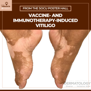
Vaccine- and Immunotherapy-Induced Vitiligo

By Posters

immunotherapy-induced vitiligo

New-onset vitiligo may be a side effect of a preventative or therapeutic medical treatment, such as a vaccine or immunotherapy. When patients on immunotherapy experience vitiligo, it’s considered a positive prognostic indicator. Cancer vaccines, biologics, immune checkpoint inhibitors, and the COVID-19 vaccine can also induce vitiligo, though what this reveals about a person’s health is unknown. In patients with darker skin tones where vitiligo may be more apparent, the development of vitiligo can have unique psychosocial impacts.

A poster presented at Skin of Color Update 2025 investigates the pathogenesis of vaccine- and immunotherapy-induced vitiligo — including the impact of these conditions on patients with skin of color — and shares management strategies for dermatology clinicians. I interviewed the poster’s lead author, Sara Omari of the William Carey University College of Osteopathic Medicine.

You investigated the factors which contribute to the pathogenesis of vitiligo induced by immunotherapies and vaccines. How did you conduct your review and what did you discover?

We reviewed a variety of studies including clinical trials, case series, systematic reviews, and other studies on the topic of immune-mediated vitiligo. We found that immunotherapies enhance antigen presentation and Th1 immune responses, leading to CD8+ T-cell-mediated destruction of melanocytes.

How common is immunotherapy-induced vitiligo (IIV)?

IIV occurs in 2-16% of melanoma patients receiving immunotherapy, but the incidence varies by the particular agent examined.

IIV is a positive outcome for melanoma patients undergoing immunotherapy treatment. How is this the case?

The depigmentation effect reflects anti-melanocyte immunologic activity. Systematic reviews have shown that melanoma-associated vitiligo in particular is associated with lower disease progression and mortality. Hence, vitiligo-like depigmentation is a favorable prognostic marker in melanoma.

How long is the onset of IIV after immunotherapy treatment has started?

The onset of vitiligo will vary by immunotherapy. Vaccine-associated cases occur within 1-3 weeks, whereas biologics may induce vitiligo within one or more months after treatment.

You also looked at how IIV presents in patients with skin of color. Did you discover any research of the sort and, if so, what did you find?

In patients with darker skin tones, vitiligo is more visually apparent, more stigmatizing, and more likely to be misdiagnosed. Commonly cited misdiagnoses included tinea versicolor or post-inflammatory hypopigmentation.

How may dermatology clinicians do a better job of treating and researching IIV, especially in patients with skin of color?

Dermatologists and other providers should inquire about new-onset hypopigmentation in their patients, use a Wood’s lamp to rule out other causes, and address stigmatization and cultural norms. Further research is necessary to unveil the ways in which IIV uniquely affects patients with skin of color.

Is there anything else a dermatology clinician should know about vaccine- and immunotherapy-induced vitiligo?

Vitiligo in the setting of immunotherapy is not necessarily a sign of treatment failure. By contrast, it may be a positive prognostic indicator. Stigma reduction during treatment is especially important when treating patients with skin of color.

Additional authors of the poster include:

Sreshta Jannu, William Carey University of Osteopathic Medicine

Mashal Zaide, William Carey University of Osteopathic Medicine

Sumra Din, William Carey University of Osteopathic Medicine 

Radhika Misra, Rowan-Virtua School of Osteopathic Medicine

Daniel N. Thompson, William Carey University College of Osteopathic Medicine

Stuti Prajapati, St. John’s Episcopal Hospital