Tag

Melasma

Challenging Cases in Skin of Color Dermatology Patients

By Media Coverage, Medical Dermatology, Sessions
Skin of Color patient dermatology cases

Source: Next Steps in Derm

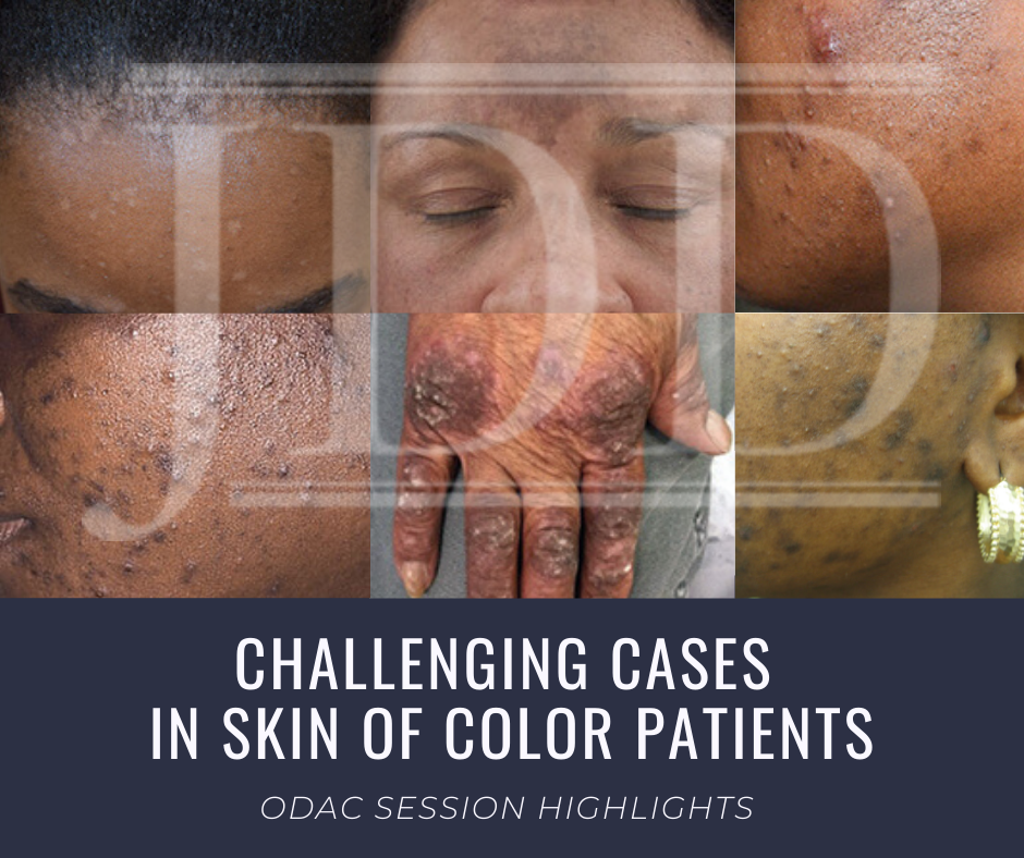
This year at the 17th Annual ODAC Dermatology, Aesthetic & Surgical Conference (ODAC), Dr Amy McMichael presented the audience with new pearls of advice on how to approach and diagnose complex medical dermatology cases in patients with skin of color. During her session, she addressed the important need for providers to be able to recognize disease in patients of all races. The majority of the global population consists of people with skin of color and the US population is changing to include a higher percentage of patients with diverse backgrounds. She covered a wide range of diagnoses from psoriasis to melasma and how these may present differently is darker skin types. As she walked the audience through each case it became apparent that being able to recognize and treat certain conditions in patients with skin of color is not only essential but also complex in nature.

First, Dr McMichael summarized the top conditions that African American patients were evaluated for during a dermatologist visit. The top 6 conditions included:

This helped to set the scene for the first case involving a 40-year-old African American female with hidradenitis suppurativa presenting with draining gluteal plaques. Even though the biopsy showed granulomatous dermatitis, the patient was not improving with multiple treatments and developed worsening pain and drainage from gluteal plaques. On a second biopsy the pathology showed psoriasis with granulomatous changes. The patient eventually improved with the systemic treatment Humira, a TNF-a inhibitor. Her major takeaways from this case included:

  • Do a second biopsy if the patient’s skin is not responding as expected to the treatment you have prescribed
  • Psoriasis can have a unique presentation similar to existing hidradenitis
  • Use systemic treatments early to help control symptoms

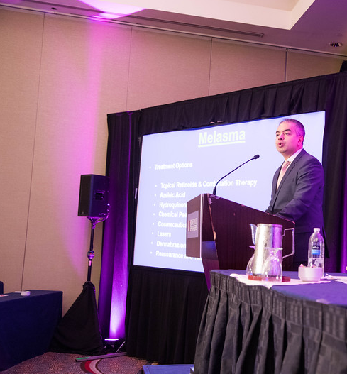
Second, she tackled the challenge of treating melasma with combination therapies. In melasma, there is too much melanin being created by melanocytes and it is then carried by keratinocytes. These cells then release melanin into the dermis, causing blotchy pigmentation often on the face. Topical therapies are usually directed towards preventing increased creation of melanin by melanocytes. People often use hydroquinone 2% or 4% along with encouragement of consistent daily sunscreen use. If used at too high of a concentration, then hydroquinone may cause ochronosis (skin becomes bluish – grey).

Dr McMichael suggested adding a novel treatment called cysteamine to the regimen for melasma treatment for more effective results. Cysteamine is an aminothiol that is made in our cells from the amino acid cysteine. Although more interest is arising now for its use in treating melasma, cysteamine was actually researched in 1966 when scientist Dr Chavin injected it into black goldfish skin and observed partial depigmentation. Cysteamine 5% cream may be a more effect treatment for melasma with less side effects.

Another novel treatment Dr McMichael discussed was the use of tranexamic acid for resistant melasma. This is another derivative of an amino acid, lysine, and it works as an anti-fibrinolytic. It has the ability to block UV-induced plasmin activity within keratinocytes. Patients would need to be screened out by their providers for a past medical history of DVT, pulmonary embolism, heart disease, and stroke before starting the oral medication. She emphasized the importance of getting a good medical history related to these conditions since tranexamic acid could increase the risk of these conditions. For patients who are able to take the medication they are expected to experience a few side effects such as mild GI upset and palpitations. This medication could provide improvement for many patients with chronic melasma who have had to struggle with this condition.

Third, in the next case we were reminded by Dr McMichael that keloids can be very disfiguring and distressful to patients. She talked about using intralesional Kenalog with contact cryotherapy as effective treatments of keloids. Other options for treatment included combining cryosurgery, intralesional Kenalog, and doxycycline. It was eye opening for the audience to hear her say we should be thinking about keloids not just as scars but tumors representing overgrowth of tissue. This paradigm shift of how we think about keloids can further shape how we think about treatment modalities for keloids as well.

Read More….

Annual Skin of Color Issue from the Journal of Drugs in Dermatology (JDD)

By SOC Manuscripts
JDD Cover Image

US Dermatologists, Dermatology Residents and Dermatology NP and PAs can create a free account on JDDonline.com to read the following Skin of Color annual issue highlights:

Patient-focused Solutions in Rosacea Management: Treatment Challenges in Special Patient Groups by Ahuva Cices MD and Andrew F. Alexis MD MPH aims to expand awareness of the impact of rosacea on QoL of patients of all ages, genders, and skin types. (CE credit is available)

Myths and Knowledge Gaps in the Aesthetic Treatment of Patients With Skin of Color, Andrew Alexis, MD et al.,  identifies knowledge gaps and myths concerning facial aesthetic treatment in individuals with SOC.

An online study was designed to survey facial aesthetic concerns, treatment priorities, and future treatment considerations in 2 companion articles, Understanding the Female Hispanic and Latino American Facial Aesthetic Patient and Understanding the Female Asian American Facial Aesthetic Patient.

In Vitro and In Vivo Efficacy and Tolerability of a Non-Hydroquinone, Multi-Action Skin Tone Correcting Cream Pearl Grimes, MD evaluates an alternative to HQ for improving skin tone.

A Survey-Based Comparison of Sun Safety Practices in a Representative Cohort of the General Public Versus Attendees of a Skin Cancer Screening examines sun-protection practices.

Topical Ozenoxacin Cream 1% for Impetigo: A Review explores the challenges of treating impetigo and growing concern of antimicrobial resistance.

Uncommon localizations of PLEVA pose a diagnostic challenge in An Atypical Presentation of PLEVA: Case Report and Review of the Literature


Skin of Color Update Co-Chair Dr. Eliot Battle Shares Insights into 2019 Faculty and Topics

By Sessions, Skin of Color Update Agenda

Skin of Color Update Co-Chair, Dr. Eliot Battle, discusses the elite faculty lineup and topics planned this year including hair loss, keloids, rosacea, acne, lasers, aesthetic treatments, skin cancer, medical dermatology, melasma, hyperpigmentation, vitiligo, inflammatory diseases and much, much more!

Skin of Color Update 2019 (previously Skin of Color Seminar Series) is the largest CE event dedicated to trending evidence-based research and new practical pearls for treating skin types III – VI. Attendees leave with critical annual updates and fresh practical pearls in skin of color dermatology.

Join us this year in New York City, September 7-8, 2019! Register today at https://skinofcolorupdate.com/registration-hotel-2019/

Seemal Desai Melasma Skin of Color Update

Melasma and PIH – Disorders of Hyperpigmentation

By Sessions, Skin of Color Update Agenda

During his talk at the 2018 Skin of Color Seminar Series (now known as the Skin of Color Update), Dr. Seemal Desai discussed the treatment of melasma (post inflammatory hyperpigmentation PIH) with a particular emphasis on new and emerging therapies. Dr. Bridget Kaufman, onsite correspondent for the meeting, shares highlights directly from the talk.  *Clinical pearls* from this session are bolded, underlined, and marked with asterisks.

Dr. Desai started by stressing the importance of having an honest and upfront conversation with your melasma patients about realistic expectations for treatment. *You must emphasize to patients that this is a chronic condition and set their expectations accordingly.*While improvement with treatment is likely, there will always be underlying pigmentary changes even despite treatment. Patients must understand from the beginning that they cannot be cured of this condition.

Dr. Desai then addressed the large vasodilatation/vascular component of melasma. Although the predominant feature of melasma is the hyperpigmented patch, a pink hue becomes apparent after treatment of the brown pigment. Historically, the redness was thought to be a side effect of Modified Kligman’s Formula, but more recent research suggests that this due to an inherent expression of vascular markers in melasma lesions. This vascular component can be treated with low fluence pulsed dye laser; oral tranexamic acid has also been helpful in Dr. Desai’s practice.

Dr. Desai then addressed the relationship between thyroid disease and melasma. There are a few studies showing thyroid disease in patients with recalcitrant melasma. *If, after optimal treatment, a patient does not improve within 3-6 months, you may want to get a free T4 and TSH to screen for thyroid disease.*

So now onto the treatment of melasma! Given that triple combination cream (TCC) is the gold standard therapy for melasma, it makes sense that Dr. Desai started with a discussion of research on this product. A randomized controlled trial of TCC in Southeast and East Asian patients with melasma found that, after 8 weeks of treatment, *TCC was superior to monotherapy with hydroquinone, although it was associated with more adverse effects.* Dr. Desai has found this to be true in his practice and, therefore, he rarely prescribes hydroquinone alone. The retinoid and steroid in TCC help with desquamation and exhibit anti-inflammatory properties that counter the upregulation of prostaglandins and tyrosinase in melasma.

For more pearls like this – register for Skin of Color Update in NYC now before it is sold out.

Read more.

Excerpt provided with permission. Originally published by Next Steps in Dermatology. All rights reserved.
Acanthosis-Nigricans-Mimicking-Melasma-boost-768x578

Acanthosis Nigricans Mimicking Melasma

By Skin of Color Update Agenda

Case Report and Pearls for Accurate Diagnosis – An Interview with the Author

During the 2018 Skin of Color Seminar Series (now the Skin of Color Update), approximately twenty posters could be seen hanging in the poster viewing area.  Each of these posters highlights novel and exciting research in skin of color. In one such poster, Dr. Monica Huynh and colleagues presented a case of acanthosis nigricans mimicking melasma.

Case Report

The patient was a 45-year-old female with a pertinent past medical history of diabetes mellitus type 2. They presented with discoloration of the cheeks for several years.  She had been treated previously with hydroquinone 4% with minimal improvement. Physical examination revealed brown-gray hyperpigmented, velvety, lichenified plaques on the lateral zygoma and hollow of the cheeks bilaterally as well as on the posterior neck.  Given the patient’s clinical presentation and past medical history of diabetes mellitus, a diagnosis of acanthosis nigricans was made. The dermatology team initiated treatment with salicylic acid cleanser twice daily, hydroquinone 4% cream twice daily, and tretinoin 0.025% cream nightly. After 8 weeks, the patient was switched to triple combination cream with improvement in facial hyperpigmentation. Due to facial dryness, the cleansing regimen was changed to salicylic acid cleanser in the morning and mandelic acid cleanser plus toner in the evening.

Read more.