Tag

Psoriasis

Pemphigus Foliaceus

Psoriasiform Pemphigus Foliaceus in an African American Female: An Important Clinical Manifestation

By Case Reports, SOC Manuscripts

JDD authors document and highlight this atypical psoriasiform presentation of Pemphigus Foliaceus in a patient with skin of color to raise awareness and improve diagnosis and outcomes.

Case Report

A 50-year-old African-American woman presented to the dermatology clinic with a pruritic eruption of 3 years’ duration that began as discrete plaques on the inframammary folds and subsequently spread towards the mid-chest, ears, back, elbows, knees, and scalp. Past treatments by other clinicians included clotrimazole cream and a topical corticosteroid of unknown potency without significant improvement. She denied any new medications and was taking aspirin, divalproex, mirtazapine, cetirizine, venlafaxine, atorvastatin, and omeprazole.

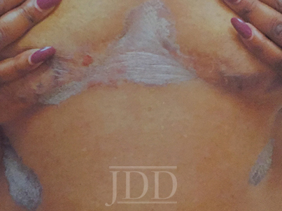
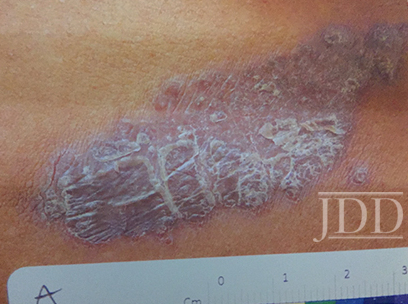
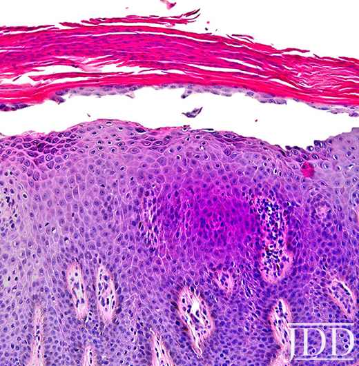
On clinical examination, the patient had well-demarcated, pink, atrophic plaques and superficial erosions over the inframammary folds and mid-chest (Figures 1). She also had well-demarcated, hyperpigmented, hyperkeratotic scaly plaques over the abdomen, suprapubic region, elbows, knees, and back with sporadic small superficial blisters (Figure 2). Complete blood count, complete metabolic panel, rheumatoid factor, and antinuclear antibody were within normal limits. Rapid plasma reagin test was negative. Erythrocyte sedimentation rate was elevated at 54 millimeter/hour (reference range 0-22 millimeter/hour). A punch biopsy of the right abdomen was performed and revealed psoriasiform epidermal hyperplasia, focal parakeratosis, and acantholysis throughout the superficial spinous and granular layers (Figure 3). Only a sparse inflammatory infiltrate was present in the underlying dermis. These clinical and histological findings supported the diagnosis of pemphigus foliaceus (PF). Patient was started on 50 mg oral dapsone daily.
Pemphigus Foliaceus

FIGURE 1. Well-demarcated, scaly plaques over the mid-chest and inframammary folds, with a single superficial erosion on the right medial chest.
Pemphigus Foliaceus

FIGURE 2. A plaque from the abdomen demonstrates “corn flake-like” scale.
Pemphigus Foliaceus

FIGURE 3. Composite photomicrograph, hematoxylin, and eosin, original magnification x200.


Discussion

Herein, we present a case of Pemphigus Foliaceus with a psoriasiform clinical presentation in an African-American patient. PF is an autoimmune skin disease caused by antibodies against the desmosomal glycoprotein, desmoglein 1.¹ Desmogleins, members of the cadherin family, serve to anchor epidermal desmosomes between adjacent keratinocytes and assist in epithelial differentiation.² Antibodies targeting desmoglein 1 result in acantholysis in the upper epidermis with limited separation in the basal layers and minimal mucosal involvement as desmoglein 1 is primarily expressed in the granular layer of the non-mucosal epidermis.¹ Patients present with scaly plaques on an erythematous base and fragile shallow blisters which are infrequently found intact; rarely, the condition can progress to exfoliative erythroderma.1,3 Initially, PF usually presents on the trunk, face, or scalp, but may subsequently involve other regions of the skin.1 Diagnosis may be confirmed with biopsy and direct immunofluorescence with intercellular IgG and C3 limited to the upper epidermis. Treatment includes oral and topical steroids, azathioprine, dapsone, and rituximab. The differential diagnosis for PF may include systemic lupus erythematosus, bullous impetigo, psoriasis, and seborrheic keratosis depending on the presentation.1

Physical exam findings in this patient were suggestive of PF due to the presence of superficial secondary erosions and “corn flake-like” scales, but psoriasis was included in the differential diagnosis due to the presence of discrete plaques with an erythematous border. PF is a relatively rare condition with a prevalence of less than 1 case per 100,000 and is about 5 to 10 times less common than pemphigus vulgaris.4 In contrast, psoriasis impacts approximately 2-4% of people in the United States.5 An endemic form of PF, fogo selvagem, has been reported in Brazil, Colombia, Peru, and Tunisia, while pemphigus vulgaris is more common in Mediterranean and Ashkenazi Jewish populations.4

We hypothesize that patients with psoriasiform presentations of PF may be misdiagnosed with plaque psoriasis. One author (JJ) has previous significant clinical experience with patients with skin of color and has seen other skin of color patients present with a psoriasiform manifestation of PF. PF and psoriasis share similar treatments including topical corticosteroids and immunosuppressants, and this may lead to underreporting of PF with psoriasiform manifestations. It is important to distinguish between these findings as there is evidence that ultraviolet light, a common treatment for psoriasis, may exacerbate PF.6,7 We performed a search of the published literature and identified one article that describes three patients with pemphigus erythematosus, a variant of PF, which was misdiagnosed as psoriasis.8 No identified articles described cases of PF with a psoriasiform presentation in patients with skin of color. We document and highlight this atypical psoriasiform presentation of PF in a patient with skin of color to raise awareness and improve diagnosis and patient outcomes.

Disclosures

The authors have no relevant disclosures. The contents do not represent the views of the U.S. Department of Veterans Affairs or the United States Government. This material is the result of work supported with resources and the use of facilities at the Sacramento VA Medical Center.


References

  1. James KA, Culton DA, Diaz LA. Diagnosis and clinical features of pemphigus foliaceus. Dermatol Clin. 2011;29(3):405-412, viii.
  2. Simpson CL, Patel DM, Green KJ. Deconstructing the skin: cytoarchitectural determinants of epidermal morphogenesis. Nat Rev Mol Cell Biol. 2011;12(9):565-580.
  3. Kershenovich R, Hodak E, Mimouni D. Diagnosis and classification of pemphigus and bullous pemphigoid. Autoimmun Rev. 2014;13(4):477-481.
  4. Meyer N, Misery L. Geoepidemiologic considerations of auto-immune pemphigus. Autoimmun Rev. 2010;9(5):A379-A382.
  5. Rachakonda TD, Schupp CW, Armstrong AW. Psoriasis prevalence among adults in the United States. J Am Acad Dermatol. 2014;70(3):512-516.
  6. Ruocco V, Ruocco E, Schiavo AL, Brunetti G, Guerrera LP, Wolf R. Pemphigus: Etiology, pathogenesis, and inducing or triggering factors: Facts and controversies. Clin Dermatol. 2013;31(4):374-381.
  7. Aghassi D, Dover JS. Pemphigus foliaceus induced by psoralen-UV-A. Arch Dermatol. 1998;134(10):1300-1301.
  8. Oktarina DA, Poot AM, Kramer D, Diercks GF, Jonkman MF, Pas HH. The IgG “lupus-band” deposition pattern of pemphigus erythematosus: association with the desmoglein 1 ectodomain as revealed by 3 cases. Arch Dermatol. 2012;148(10):1173-1178.

Originally published in the Journal of Drugs in Dermatology in April 2018. 

Evan Austin BS, Jillian W. Millsop MD, Haines Ely MD, Jared Jagdeo MD MS, and Joshua M. Schulman MD (2018). Psoriasiform Pemphigus Foliaceus in an African American Female: An Important Clinical Manifestation. Journal of Drugs in Dermatology, 17(14), 471-473. https://jddonline.com/articles/dermatology/S1545961618P0471X 

Content and images republished with permission from the Journal of Drugs in Dermatology.

Adapted from original article for length and style.

The Journal of Drugs in Dermatology is available complimentary to US dermatologists, US dermatology residents, and US dermatology NP/PA. Create an account on JDDonline.com and access over 15 years of PubMed/MEDLINE archived content.

Did you enjoy this case report? You can find more here.

Challenging Cases in Skin of Color Dermatology Patients

By Media Coverage, Medical Dermatology, Sessions
Skin of Color patient dermatology cases

Source: Next Steps in Derm

This year at the 17th Annual ODAC Dermatology, Aesthetic & Surgical Conference (ODAC), Dr Amy McMichael presented the audience with new pearls of advice on how to approach and diagnose complex medical dermatology cases in patients with skin of color. During her session, she addressed the important need for providers to be able to recognize disease in patients of all races. The majority of the global population consists of people with skin of color and the US population is changing to include a higher percentage of patients with diverse backgrounds. She covered a wide range of diagnoses from psoriasis to melasma and how these may present differently is darker skin types. As she walked the audience through each case it became apparent that being able to recognize and treat certain conditions in patients with skin of color is not only essential but also complex in nature.

First, Dr McMichael summarized the top conditions that African American patients were evaluated for during a dermatologist visit. The top 6 conditions included:

This helped to set the scene for the first case involving a 40-year-old African American female with hidradenitis suppurativa presenting with draining gluteal plaques. Even though the biopsy showed granulomatous dermatitis, the patient was not improving with multiple treatments and developed worsening pain and drainage from gluteal plaques. On a second biopsy the pathology showed psoriasis with granulomatous changes. The patient eventually improved with the systemic treatment Humira, a TNF-a inhibitor. Her major takeaways from this case included:

  • Do a second biopsy if the patient’s skin is not responding as expected to the treatment you have prescribed
  • Psoriasis can have a unique presentation similar to existing hidradenitis
  • Use systemic treatments early to help control symptoms

Second, she tackled the challenge of treating melasma with combination therapies. In melasma, there is too much melanin being created by melanocytes and it is then carried by keratinocytes. These cells then release melanin into the dermis, causing blotchy pigmentation often on the face. Topical therapies are usually directed towards preventing increased creation of melanin by melanocytes. People often use hydroquinone 2% or 4% along with encouragement of consistent daily sunscreen use. If used at too high of a concentration, then hydroquinone may cause ochronosis (skin becomes bluish – grey).

Dr McMichael suggested adding a novel treatment called cysteamine to the regimen for melasma treatment for more effective results. Cysteamine is an aminothiol that is made in our cells from the amino acid cysteine. Although more interest is arising now for its use in treating melasma, cysteamine was actually researched in 1966 when scientist Dr Chavin injected it into black goldfish skin and observed partial depigmentation. Cysteamine 5% cream may be a more effect treatment for melasma with less side effects.

Another novel treatment Dr McMichael discussed was the use of tranexamic acid for resistant melasma. This is another derivative of an amino acid, lysine, and it works as an anti-fibrinolytic. It has the ability to block UV-induced plasmin activity within keratinocytes. Patients would need to be screened out by their providers for a past medical history of DVT, pulmonary embolism, heart disease, and stroke before starting the oral medication. She emphasized the importance of getting a good medical history related to these conditions since tranexamic acid could increase the risk of these conditions. For patients who are able to take the medication they are expected to experience a few side effects such as mild GI upset and palpitations. This medication could provide improvement for many patients with chronic melasma who have had to struggle with this condition.

Third, in the next case we were reminded by Dr McMichael that keloids can be very disfiguring and distressful to patients. She talked about using intralesional Kenalog with contact cryotherapy as effective treatments of keloids. Other options for treatment included combining cryosurgery, intralesional Kenalog, and doxycycline. It was eye opening for the audience to hear her say we should be thinking about keloids not just as scars but tumors representing overgrowth of tissue. This paradigm shift of how we think about keloids can further shape how we think about treatment modalities for keloids as well.

Read More….

Skin of Color Update Co-Chair Dr. Eliot Battle Shares Insights into 2019 Faculty and Topics

By Sessions, Skin of Color Update Agenda

Skin of Color Update Co-Chair, Dr. Eliot Battle, discusses the elite faculty lineup and topics planned this year including hair loss, keloids, rosacea, acne, lasers, aesthetic treatments, skin cancer, medical dermatology, melasma, hyperpigmentation, vitiligo, inflammatory diseases and much, much more!

Skin of Color Update 2019 (previously Skin of Color Seminar Series) is the largest CE event dedicated to trending evidence-based research and new practical pearls for treating skin types III – VI. Attendees leave with critical annual updates and fresh practical pearls in skin of color dermatology.

Join us this year in New York City, September 7-8, 2019! Register today at https://skinofcolorupdate.com/registration-hotel-2019/

Co-Chair Dr. Alexis Shares the Exciting 2019 Program Highlights

By Sessions, Skin of Color Update Agenda

Skin of Color Update 2019 (previously Skin of Color Seminar Series) is the largest CE event dedicated to trending evidence-based research and new practical pearls for treating skin types III – VI. Attendees leave with critical annual updates and fresh practical pearls in skin of color dermatology. Earn CE in New York City with direct access to elite experts and an experience unmatched by any other event in dermatology.

Psoriasis Skin of Color Alexis

Psoriasis in Skin of Color: Are there differences in Presentation and Treatment?

By Sessions

While previously thought to be a rare diagnosis in non-white racial ethnic groups, more recent data has shown that psoriasis in patients with skin of color is much more common than was once thought. Dr. Andrew Alexis spoke during the Skin of Color Seminar Series (now Skin of Color Update) in New York City on May 5th, 2018 on this condition and on the important differences in clinical characteristics, diagnosis, and treatment of psoriasis in skin of color.

Beginning with the diagnosis of psoriasis in skin of color, it is important to be aware that the classic red plaque typically seen in Caucasian skin may appear more violaceous or even red-brown in our skin of color patients.  This can be confusing with the similar appearing, but histologically unique entity, lichen planus. Pay close attention to the distribution of the plaques—extensor surfaces and asymptomatic will favor psoriasis whereas flexural and pruritic may favor lichen planus. Other conditions that may be more common in our skin of color patients such as sarcoidosis or hypertrophic discoid lupus also need to be considered.

Once a diagnosis of psoriasis is established, it is important to understand the intricacies of treating the psoriasis in skin of color patients. For example, when treating psoriasis of the scalp, it is important to select practices and treatments that are compatible with the patients’ hair care regimen and hair texture. It is critical to determine how frequently patients are washing their hair and how feasible it is for patients to use a daily solution or medication on the scalp. One potential option shown to have clinical benefit is a mixture of calcipotriene and betamethasone dipropionate in a castor oil vehicle.

Read more.


Excerpt provided with permission. Originally published by Next Steps in Dermatology. All rights reserved.

Treating Scalp Psoriasis in Women of African Descent

By Sessions, Skin of Color Update Agenda

Dr. Andrew Alexis is the Chair of the Department of Dermatology at Mount Sinai St. Luke’s and Mount Sinai Roosevelt. He is also Associate Professor of Dermatology at the Icahn School of Medicine at Mount Sinai. Dr. Alexis is the Co-Chair of the Skin of Color Seminar Series in New York City. During the 2017 conference he provided practical pearls and treatment outlines for African American patients with scalp psoriasis.

Dr. Alexis recommends selecting a treatment regimen that is compatible with the patient’s hair care practices including less frequent hair washing in women of African descent (typically once per week to once every other week). Daily hair washing, especially with most prescription shampoos, is often associated with increased hair dryness and breakage. In addition, it is also very time consuming for most women of African descent due to common styling practices.

Suggested Regimen for African-American Females:

  • Once weekly washing with prescription shampoo. This may be increased to two times a week depending on the severity and patient preferences
  • Continue with usual conditioner
  • Once weekly topical fluocinolone acetonide in peanut oil vehicle applied to the scalp for 6-8 hours overnight prior to washing or several times per week without washing
  • Once to twice daily application of POTENT topical steroid in vehicle that is compatible with hair care practices and hair texture (e.g. lotion, emollient foam, oil > gel, solution, ethanolic foam) Ask the patient for vehicle preferences
  • Alternative: calcipotriene and betamethasone dipropionate topical suspension daily

Attendees at the Skin of Color Seminar Series May 5-6 in NYC will have the opportunity to dig deeper into psoriasis treatment in skin of color patients as Dr. Alexis gives his latest updates, pearls and therapeutic insights and also personally answers attendees’ most pressing questions.