Tag

dermatology

SKIN OF COLOR UPDATE PRE-CONFERENCE VIRTUAL SYMPOSIUM

By Medical Dermatology, Sessions, Skin of Color Update Agenda
A Case-Based Conversation with The Experts: Treating Pigmentary Disorders in Skin of Color Patients
SKIN OF COLOR UPDATE PRE-CONFERNECE VIRTUAL SYMPOSIUM
TUESDAY, AUGUST 3RD | 6:00PM ET – 9:00PM ET
The Skin of Color Update invites you to join its pre-conference symposium where co-chairs Drs. Andrew Alexis and Eliot Battle will host an interactive, case-based conversation with pigmentary disorders experts. Faculty will review treatment options for common as well as challenging and less frequently discussed pigmentary conditions in skin of color patients. Through a detailed review of each case, panelists will provide guidance and evidence-based treatment protocols as well as practical pearls drawn from their clinical experience. You will walk away from this session armed with clinical pearls immediately useful in your practice. In addition, all panelists will participate in live Q&A sessions to answer your most pressing questions about treating pigmentary disorders in SOC.
AGENDA
6:00-6:05 PM – Welcome & Introductions from Symposium Moderators – Andrew F. Alexis, MD, MPH & Elliot F. Battle, MD
6:05-6:20 PM – A Challenging Case of Melasma – Heather Woolery-Lloyd, MD
6:20-6:35 PM – A Case of Vitiligo Treated with Pulsed Corticosteroids/JAK-Inhibitor – Seemal Desai, MD
6:35-6:50 PM – Post-Inflammatory Hyperpigmentation(PIH) Topical & Procedural Treatment – Neelam Vashi, MD
6:50-7:00 PM – Live Audience Q&A
7:00-8:00PM – For Patients with Plaque Psoriasis: An Oral, Non-Biologic Therapy With Data on Clearer Skin and Symptoms – Paul Wallace, MD, MPA (Non-CE Workshop)
8:00-8:05 PM – Welcome & Introductions from Symposium Moderators – Andrew F. Alexis, MD, MPH & Elliot F. Battle, MD
8:05-8:20 PM – A Challenging Case of Erythema Dyschromicum Perstans – Nada Elbuluk, MD, MSc
8:20-8:35 PM – A Case of Hypopigmented Mycosis Fungoides – Eva Kerby, MD
8:35-8:50 PM – Lichen Planus Pigmentosus – Mukta Sackdev, MD
8:50-9:00 PM – Live Audience Q&A
SYMPOSIUM CO-CHARIS
Andrew F. Alexis, MD, MPH
Eliot F. Battle, MD
EXPERT FACULTY
Seemal R. Desai, MD, FAAD
Nada Elbuluk MD, MSc
Eva Kerby, MD
Heather Woolery-Lloyd, MD
Mukta Sachdev, MD
Neelam Vashi, MD

The Relevance of Vitamin D Supplementation for People of Color in the Era of COVID-19

By COVID-19 Resources, Skin of Color Update Agenda
Vitamin D

Source: JDD Online

The Journal of Drugs in Dermatology recently featured the article, The Relevance of Vitamin D Supplementation for People of Color in the Era of COVID-19, authored by Skin of Color Virtual Update faculty, Pearl E. Grimes MD, and Andrew F. Alexis MD MPH along with Nada Elbuluk MD MSU.

Introduction

African Americans (AA) and other people of color are dying at highly disproportionate rates from COVID-19. The statistics are staggering: in New York City alone, per 100,000 population, death rates in AA were 92.3, and in Hispanics 74.3, compared to 45.2 in Whites and 34.5 in Asians.1 Similar numbers have been reported in other cities and are presumed underestimations, given limited racial/ethnic reporting. In the states currently releasing the number of COVID-19 deaths by race and ethnicity, Blacks make up roughly 13 percent of the population, but 27 percent of the deaths. According to the American Public Media Research Lab, the rate of COVID-19 deaths nationally for Blacks has been reported as twice the rate of deaths of Asians and Latinos in the US and more than 2.5 times the rate for White residents.

Socio-economic reasons, pre-existing comorbidities, work circumstances, inconsistent healthcare access, stress, and decreased immunity, amongst other factors, have been posited as reasons for this shocking disparity. People of color, in particular AA and Hispanics, are more likely to be uninsured and to be frontline workers during the COVID-19 pandemic. This is compounded by the fact that comorbidities such as hypertension, diabetes, asthma, obesity, and cardiovascular disease are more common in AA and are also associated with higher COVID-19 mortality rates. Emerging evidence suggests that Vitamin D deficiency may represent another risk factor for poor outcomes from COVID-19.

Relevance of Vitamin D
Vitamin D is a secosteroid hormone synthesized in the skin following exposure to UVB ultraviolet radiation where it mediates the conversion of 7-dehydrocholesterol to pre-Vitamin D3. Following transport to the liver, it is hydroxylated to 25(OH)D, the primary circulating form typically used to measure serum Vitamin D levels. 25(OH)D is subsequently converted to the biologically active form 1,25, dihydroxy vitamin D in the kidneys by 1-alpha hydrolase. This active form binds to its nuclear Vitamin D receptor to induce the transcription of over 200 genes, affecting a wide range of physiologic functions.

Multiple studies have documented significant Vitamin D deficiency in people of color, especially in AA. Heavily melanized skin retards the synthesis of Vitamin D and necessitates longer periods of sun exposure for adequate synthesis of Vitamin D. Ginde et al. assessed demographic differences and trends of Vitamin D insufficiency in a US population.2Serum 25(OH)D levels were compared over two time periods (1988–1994 and 2001–2004) from the Third National Health and Nutrition Examination Survey (NHANES III) data base including two large populations (n=18,883 and n=13,369, respectively). Non-Hispanic Blacks had a significantly higher prevalence of Vitamin D deficiency, increasing in severity in the later data base. Recent NHANES data from 2011–2014 further documented the high risk of deficiency in non-Hispanic Blacks. In a recent prospective cohort study of 14,319 subjects, an estimated 65.4% of non-Hispanic Blacks were deficient in Vitamin D, compared to 29% of Hispanics and 14% of non-Hispanic Whites.3

Vitamin D deficiency has been shown to be a risk factor for many of the comorbidities that disproportionately plague AA including diabetes, hypertension, cardiovascular disease, autoimmune diseases such as lupus erythematosus, as well as aggressive forms of breast and prostate cancer.4 While the classic role of Vitamin D involves calcium and phosphorus homeostasis for healthy bone metabolism, it exerts a spectrum of pleotropic effects impacting cell growth, differentiation, inflammation, and immune regulation. Healthy levels of Vitamin D have been linked to significantly reduced mortality and improved health outcomes. Numerous investigations document the prolific role of Vitamin D in antimicrobial defense and modulation of the innate and adaptive immune responses. It mediates the induction of key antimicrobial peptides in the respiratory epithelium including cathelicidin (LL37) and beta defensins, which destroy invading organisms. In addition, Vitamin D inhibits the production of pro-inflammatory cytokines including IL-2, IFN-γ, TNF-α, and IL-6, while promoting Th2 responses by increasing IL-4, IL-5, and IL-10 production, hence skewing T cell responses to a down regulated, anti-inflammatory state.4

For the general population, the US Institutes of Medicine (IOM) recommends Vitamin D supplementation at doses that vary according to age and are based primarily on bone health. Current IOM supplementation recommendations are 400 IU (10ug) for infants, 600 IU/d (15ug) for children, adolescents, and adults, and 800 IU/d (20ug) for adults aged over 70 years to maintain a 25(OH)D concentration of 20ng/mL or higher. However, in individuals who are deficient in Vitamin D (25(OH)D level <20 ng/ mL), of which patients with skin of color are at a higher risk, supplementation is considerably higher. These recommendations are summarized summarized in Table 1.5

Conclusions

Vitamin D deficiency has been well documented in people of color, in particular AA. The aforementioned data suggest a relationship between low Vitamin D status and COVID-19 mortality rates. While myriad socioeconomic and health care disparities may be contributing factors, we must indeed consider key biological variables, including Vitamin D status, that may impact these observations. Future prospective studies are necessary to confirm these findings. As there is currently no readily available treatment or vaccine for COVID-19, treating physicians should be cognizant of the higher prevalence of Vitamin D deficiency in skin of color populations and its emerging potential role in COVID-19 outcomes. Given the devastating statistics of COVID-19 among minority communities and the multifaceted role of Vitamin D in skin and systemic health, dermatologists are essential partners in decreasing health care disparities by initiating the vitamin D dialogue. As such, we can play an invaluable role in improving the health outcomes of our patients, particularly people of color, during and beyond the COVID-19 pandemic.

Read Full Article….

Frontal Fibrosing Alopecia Presenting as Androgenetic Alopecia in an African American Woman

By Aesthetic Dermatology, Medical Dermatology, Sessions, Skin of Color Update Agenda
Alopecia patient picture

Source: Next Steps in Derm

Frontal fibrosing alopecia (FFA) is a primary lymphocytic cicatricial alopecia that is currently regarded as a variant of lichen planopilaris. FFA has historically been considered rare in black patients, in whom traction alopecia, central centrifugal cicatricial alopecia, and androgenetic alopecia are frequently assumed to be more common. JDD author Kimberly Huerth, MD, ME describes a case of FFA in a black woman that both clinically resembled androgenetic alopecia and lacked many of the physical exam and dermoscopic findings associated with FFA. In doing so, she highlights the need for physicians to have a high index of suspicion for FFA in any black patient who presents with frontotemporal alopecia.

REPORT OF A CASE

A 53-year-old African American woman presented with a 6-month history of asymptomatic, moderately severe hair loss along the frontal hairline, which had not stabilized or improved with minoxidil 2% solution BID. Physical exam revealed decreased hair density affecting the frontal scalp, suggestive of androgenetic alopecia (Figure1). Dermoscopic examination showed decreased follicular ostia without perifollicular scaling or erythema. Eyebrow alopecia, facial papules, and glabellar red dots were absent, and there was no associated loss of body hair. A 4-mm punch biopsy sent for histopathologic examination revealed dense, chronic, perifollicular inflammation affecting the mid and upper portions of the follicles, with loss of associated sebaceous glands. Involved hairs demonstrated vacuolar interface disruption of the basilar and epibasilar layers at the level of the isthmus and infundibulum, with prominent exocytosis of lymphocytes into the outer root sheath. There was no miniaturization, dermal mucin, or inflammation affecting the epidermis, arrector pili muscles, and eccrine glands (Figure 2).

A diagnosis of FFA was confirmed by these findings. Our patient was managed with intralesional triamcinolone acetonide (10mg/cc) injections, clobetasol 0.05% ointment BID, hydroxychloroquine 200 mg PO BID, and minoxidil 5 mg PO daily. Unfortunately, her alopecia did not stabilize with these measures.

DISCUSSION

FFA is a primary lymphocytic cicatricial alopecia that is currently regarded as a variant of lichen planopilaris. It is characterized by band-like frontotemporal hairline recession, often with associated eyebrow alopecia, perifollicular erythema, and scaling. Clinical findings are frequently accompanied by pruritus and burning of the affected scalp. Since it was first described in 1994,1 FFA has largely been viewed as an alopecia of post-menopausal Caucasian women. This archetype has been maintained by patient demographics of subsequent published case series.2,3 FFA may thus be underdiagnosed in black women, in whom traction alopecia, central centrifugal cicatricial alopecia, and androgenetic alopecia are assumed to be more common. Furthermore, FFA can manifest uniquely in black women, who may be premenopausal4,5 and asymptomatic4 at the time of presentation. Classic signs of FFA may be subtle or absent among black patients, as increased pigmentation may render erythema difficult to appreciate, while oils and hair care products may diminish the appearance of scale.

It is important for dermatologists to both recognize that FFA is not uncommon in the black population,4,5 and to acknowledge how it initially came to be regarded as a disease of post-menopausal white women. Several of the larger published series come from geographic areas that lack a substantial skin of color population.2,3 There are also socioeconomic factors to consider. One series comprised exclusively of Caucasian women found their patients to be more affluent, which was speculated to be a surrogate marker for an unknown risk factor associated with the development of FFA.3 What these authors did not discuss, however, is that affluence enables access to specialty medical care. Affluence affects insurance status, which has been shown to vary widely among racial groups.6 Insurance status in turn bears upon who has access to dermatologic care, and who is ultimately included in a case series.

Read more…..

The Business of Dermatology

By Aesthetic Dermatology, Medical Dermatology
The Business of Dermatology

Business intellect, a vital aspect of managing a practice, is not taught in residency. From the infancy of their training, dermatologists are trained to think broadly and scrupulously, using each clue, each corporeal sense, and each available tool to accurately diagnose and manage a plethora of cutaneous conditions. After residency, dermatologists set out armed with the knowledge and drive to deliver expert care to their future patients. However, despite their education and best intentions, lack of business acumen can hinder even the brightest and most motivated of practitioners. In order to enlighten oneself in the complicated field of business management, clinicians are left to fend for themselves, often learning as they go, sometimes making unnecessary mistakes, and adjusting their business practices reactively. Retrospective “trial and error” learning is time-consuming, cumbersome, and costly. Why not short track and get the goods without the trial and error, making costly mistakes and taking years. The new book, The Business of Dermatology is a cornerstone achievement in the standardization of business education for dermatologists.

Edited by Drs. Jeffrey S. Dover and Kavita Mariwalla, and authored by impressive experts in the field, The Business of Dermatology offers a comprehensive guide to opening, maintaining, and sustaining a practice. To start, the power of this textbook fundamentally lies in the experience and scope of its authorship. The authors were hand-selected by the editors ensuring that each chapter was written by a tried and true expert in that subject. Unlike other textbooks in the field of business management and administration that are primarily written by individuals from the business world, some of whom have no insight into the inner machinations of the medical world, or hands-on experience, the authors of this book are well-known, respected dermatologists that hail from thriving practices of their own. The reader has an unprecedented opportunity to learn from the firsthand experiences of top authorities who live and breathe dermatology. Using conversational prose, the authors depict their experiences, trials, and errors, employing specific real-world examples and scenarios while tackling each subject.

Read More….

Recommendations for Managing Lichen Planopilaris With Hydroxychloroquine During the COVID-19 Pandemic

By COVID-19 Resources
Recommendations Chart

Source: Journal of Drugs in Dermatology

The recommendations are noted in the article, Considerations of Managing Lichen Planopilaris With Hydroxychloroquine During the COVID-19 Pandemic, will be available in the June print issue of the Journal of Drugs in Dermatology.

Chloroquine (CQ) and hydroxychloroquine (HCQ), two well-known drugs among dermatologists, have shown their efficacy in the inhibition of severe acute respiratory syndrome coronavirus 2 (SARS-CoV-2) replication.1,2 HCQ is found to possess a better clinical safety profile, more potency, and fewer drug–drug interactions compared to chloroquine.3 HCQ has been reported to exert efficacy in the inhibition of SARS-CoV-2 in vitro replication through diverse mechanisms. First, it interferes with the glycosylation of angiotensin-converting enzyme 2 (ACE2), resulting in a subsequent reduction in the binding efficacy between ACE2 on host cells and the SARS-CoV-2 spike protein. Second, it blocks the fusion of the virus to the host cell. Finally, it suppresses the “cytokine storm” accountable for the disease progression to acute respiratory distress syndrome (ARDS). Although studies are underway to confirm the in vivo effectiveness of HCQ in the SARS-CoV-2 infection, promising primary results have led to a shortage of the drug for dermatologic purposes, which is a real concern in the current pandemic.1

While we are amid a pandemic with the possible shortage of HCQ, dermatologists should be reminded that:

  • The anti-inflammatory effect of HCQ may improve the clinical signs of LPP; however, administration of this drug is insufficient to prevent the subclinical disease progression.9 Dermatologists may discontinue the use of HCQ in responders after 1 year with monitoring the patients for recurrence or relapse.5
  • Topical and intralesional super potent corticosteroids are recommended as the first-line treatment in localized LPP.4
  • Oral cyclosporine followed by systemic corticosteroid may be the most effective medications in LPP; however, disease relapse may be detected.10 Mycophenolate mofetil has a more favorable safety profile compared to cyclosporine11 but the immunosuppressive nature of these medications necessitates extreme caution toward their administration during COVID-19 pandemic.12
  • Acitretin (25 mg/day) may be an appropriate alternative since it has shown improvement in 66% of patients.7
  • Pioglitazone (hypoglycemic drug, 15–30 mg/day) has shown some efficacy in the treatment of LPP and can be considered as an alternative to HCQ.4
  • Tetracyclines antibiotics can also be considered as an alternative due to favorable outcomes in previous studies.13

Read Full Article….

Ethnicity Matters: Medical Dermatology Concerns Across Ethnic Groups

By Medical Dermatology, Sessions
Wendy Roberts Presenting at SOCU

Source: Dermatology News

This is an excerpt from Dermatology News’ coverage of Skin of Color Update 2019.

For women with pseudofolliculitis barbae, an empirically-based strategy of microdermabrasion, laser treatment, emollients, and maintenance retinoids has been found highly effective, Wendy Roberts, MD, reported at the Skin of Color Update 2019.

“We didn’t have great treatments for this problem in the past, but the technology has evolved, and you can now get most women clear,” Dr. Roberts, a dermatologist who practices in Rancho Mirage, Calif., said at the meeting.

This approach is appropriate in all women, but Dr. Roberts focused on her experience with black patients, for whom an antioxidant cream is added to address the inflammatory-associated hyperpigmentation that often accompanies pseudofolliculitis barbae, a chronic inflammatory skin condition typically characterized by small, painful papules and pustules.

Start with microdermabrasion to treat the hypertrophic hair follicles and address keratin plugs, Dr. Roberts said. The microdermabrasion smooths the skin and increases penetration of subsequent creams and topics, she said.

“In the same session, I treat with Nd-YAG 1064 nm laser using short pulses,” she noted. For black women, she makes four passes with the laser at a level of moderate intensity. For those with lighter skin, she might perform as many as six passes with the laser set higher.

The microdermabrasion is repeated monthly for three or four treatments, but can be extended for those with persistent symptoms, Dr. Roberts pointed out. She presented a case of a patient who required seven treatments to achieve a satisfactory response.

Read More….

On-Demand Recording: COVID-19: Special Considerations for the Skin of Color Patient – A Conversation with the Experts

By COVID-19 Resources

This webinar was previously recorded on April 27th, 2020 and is now available on demand. Almirall has graciously supported the on-demand broadcast of this webinar.

Skin of Color Update (SOCU) and JDD invite you to attend “COVID-19: Special Considerations for the Skin of Color Patient – A Conversation with the Experts”. During this 90-minute webinar, Dr. Andrew Alexis will be joined by skin of color key opinion leaders to discuss special considerations and practical approaches to managing dermatologic disorders in skin of color during the COVID-19 pandemic.


Topics to be addressed include the following:

  • Hair Disorders
  • Pigmentary Disorders
  • Aesthetic Concerns
  • Inflammatory Disorders (including acne, atopic dermatitis, psoriasis, and others)
  • How to stay connected with your skin of color patients on social media
  • Vitamin D deficiency among people of color and its potential relevance to COVID-19 in patients of color
  • COVID-19 related shifts in common dermatologic concerns in skin of color
  • Impact on the practice of aesthetic dermatology for skin of color

MODERATOR:

Andrew Alexis, MD, MPH (Chair, Department of Dermatology, Mount Sinai West and Mount Sinai Morningside and Professor, Icahn School of Medicine at Mount Sinai)


PANELISTS:

Eliot F. Battle, Jr., MD (CEO and Co-Founder of Cultura Dermatology & Plastic Surgery)

Amy McMichael, MD (Professor and Chair of Dermatology, Wake Forest Baptist Medical Center)

Heather Woolery-Lloyd, MD (Director, Skin of Color Division, University of Miami School of Medicine)

Pearl Grimes, MD (Founder and Director, Vitiligo and Pigmentation Institute of Southern California)

Supported by:

Almirall Logo

Long-Term Benefits of Daily Photo-Protection With a Broad-Spectrum Sunscreen in United States Hispanic Female Population

By Aesthetic Dermatology, Medical Dermatology
Image of photo aging

Source: JDD Online

The following is an excerpt from the Journal of Drugs in Dermatology article, Long-Term Benefits of Daily Photo-Protection With a Broad-Spectrum Sunscreen in United States Hispanic Female Population.

Introduction
The demographics of the United states are evolving with a large increase in racial and ethnic diversity driven by international migration of Hispanic, African, and Asian populations leading to a minority-majority shift in ~2050 towards persons of color (Fitzpatrick III, IV, V, and VI).1 Specifically, the Hispanic population is projected to be among the fastest growing population in the US, projected to increase from 55 million in 2014 to 119 million in 2060, a change of +115%.1

Subjects with skin of color are heterogeneous with multiple shades and tones and different reactions to intrinsic and extrinsic aging factors due to structural and physiologic differences.2,3 Skin of color individuals have fewer visible signs of aging (deep wrinkles, fine lines, rough surface texture, and sun spots). However, darker skin tones are more susceptible to certain skin conditions including post-inflammatory hyperpigmentation (may occur after acne, eczema, injury, laceration, melasma, post-inflammatory hypopigmentation, pityriasis alba (round, light patches covered with fine scales), dry or “ashy” skin, dermatosis papulosa nigra, and/or greater risk of keloid development.2,3 The incidence of skin cancer among US Hispanics has also increased 1.3% annually from 1992 to 2008.4

Photodamage is characterized histologically by degeneration of the connective tissue and abnormalities in keratinocytes and melanocytes. Clinically, it manifests primarily with wrinkles, dyschromia, texture changes, and, in more severe cases skin cancer.5 Formulations containing broad spectrum sunscreens against both UVA and UVB play an essential role in the prevention of photodamage and UV-induced skin cancers.6,7,8 However, the majority of clinical research on photoprotection has been conducted on subjects with Fitzpatrick types I to III skin and have reported improvements in signs associated with skin aging and texture.9,10 Verschoore et al was the first to conduct a short-term clinical study in India with Phototype IV and VI subjects, and provided first evidence on the effectiveness of daily sunscreen use on skin tone and radiance.11 Similar benefits were observed in an 8-week study in US.12

Although sun protection is highly recommended by dermatologists for skin cancer risk-reduction and the prevention of premature aging or pigmentary disorders, adherence to the recommendations is not commonly observed among US Hispanics.13 Moreover, a large number of US Hispanics reside in areas with high UV index with a high degree of sun seeking behavior. Among Hispanic adults who report engaging in sun protection, they do so mostly by staying in the shade (53.7%) rather than use of sunscreen (32.3%) or wearing sun protective clothes (18.1%); while 36.7% of the subjects surveyed indicated that they never use sunscreen.14,15 There are sociodemographic factors that contribute to the adherence to safe sun behaviour such as education, age, and gender, etc, therefore there is a need to raise awareness of skin cancer risks, advocate for preventive measures and educate on benefits of sunscreen and sun protection among US Hispanics.16

The benefits of topical agents for reversal of sun damage has been well established. Use of retinoic acid and its derivatives or other drugs to reverse and improve sun damaged skin has been demonstrated in many studies.17,18 Long-term sunscreenuse along with other topical agents have also been shown to prevent photodamage and hyperpigmentation in fair-skinned subjects.19 For effective photoprotection, sunscreen products containing both SPF and PPD are essential to battle the harmful UVB (skin cancer risks) and UVA (photo-aging risks).20 Daily use of a broad-spectrum sunscreen (SPF 30) over a one-year period has also been demonstrated to improve clinical parameters of photodamage in phototype I-III subjects.10 However, a comprehensive long-term sunscreen use study in skin of color is lacking. Therefore, this study was designed to assess the benefits of sunscreen of SPF30/PPD 20 in Hispanic women of Fitzpatrick skin types IV and V over 12 months in comparison to a real-life observational group with subjects who did not use sunscreen regularly.

Read Full Article….

Discussions and Conclusions
Effective photoprotection is critical for healthy skin, in preventing skin cancers, reducing photodamage, and improving aesthetic appearance. A broad spectrum sunscreen protecting against both UVA and UVB irradiation is essential. Protecting against the UVA spectrum needs special attention, especially under daily diffused exposure, as UVA is more penetrating and less affected by seasonality and impacts photoaging and skin oxidative stress.22 It has been reported that in order to receive effective photoprotection on skin, a PPD value of 18 is desired.20 In this study, the investigational product with SPF 30/PPD 20 is considered sufficient for daily activity without prolonged direct sun exposure when applied properly. Concerning skin of color population, the use of sunscreen is lower than in Caucasians despite high prevalence of sun-related pigmentary disorders and rising rates of cutaneous cancers.4 This study provides strong evidence to educate and advocate for daily use of a proper sunscreen product for populations with high phototype skin.

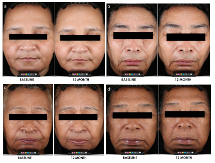
The clinical evaluation demonstrated significant visible improvement in sunscreen group starting from 3 months and progressive increased over time. Benefits on multiple facial areas and body sites were visible (upper, mid- and lower face, neck, and hands), not only on pigmentary-related concerns (skin tone evenness, overall hyperpigmentation, dark spots, and blotchiness), but also on aging parameters such as fine lines, skin texture, and overall skin quality. This suggests that beyond the preventative benefits, long-term persistent use of a proper sunscreen may also allow the photodamaged skin to self-heal and repair over time.

Histological observations further supported the clinical findings. The observation that the real-life group had higher tendency for pigmentation incontinence is of strong research interest. It has been reported that UV irradiation can destabilize and damage the dermal-epidermal junction (DEJ), which facilitates the entrapment of melanin in the dermis.23 The dermal melanin is extremely difficult to remove, often resulting in stubborn hyperpigmentation.24 This is especially important for skin of color population in whom dermal hyperpigmentation lesions are common and can be worsened with excessive sun exposure. This study provides the first evidence that effective daily photoprotection can be a strategy to prevent dermal melanin formation by protecting the DEJ. A larger sample size study with DEJ biomarkers will help to further elucidate this hypothesis. Infiltration of CD68-positive Macrophages is a hallmark of the inflammatory response after UV irradiation. In the dermis, 2 out of 3 of the real-life biopsy samples showed significant increase in CD68 positive macrophage cells at 12 months compared to baseline, while such change was not observed in the sunscreen group. This suggests the potential preventative benefits of sunscreen in subclinical skin inflammation induced by chronic exposure to UV. In all of the histological evaluations, thegeographical location in which the study was conducted (Los Angeles versus Washington, DC) was not a strong contributing factor to any of the observed differences. However, the histological findings in this study are limited by the small number of biopsies obtained.

In summary, this 12-month study on long-term persistent use of an SPF30/PPD20 sunscreen on phototype IV and V subjects demonstrated significant improvement in skin quality and improvement in skin color and photoaging parameters. To our knowledge, this is the first study of this kind in skin of color and Hispanic population. This study confirms that effective sunscreen use is not only protective and beneficial for light skin population but is also critical in improving skin condition for skin of color patients. Overall, the study demonstrates that daily use of sunscreen can protect skin from photo related damage and even reverse some of the photo-damage that has already occurred in skin. In addition to previous studies that demonstrated the photo-protective properties of sunscreen use in normal and diseased skin states7,8,9,10 and in view of the fact that good photoprotection behaviors are not common among Hispanics,14,15,16 studies of this type can help educate and stress the importance of daily use of sunscreen and other sun protection behaviors in Hispanic and other skin of color populations.

Read Full Article….

Intralesional Triamcinolone Acetonide in the Treatment of Traction Alopecia

By Aesthetic Dermatology, Case Reports, Medical Dermatology, Sessions
Patient with Alopecia

Source: Next Steps in Derm

In this case series, JDD authors evaluate the efficacy and safety of intralesional triamcinolone acetonide injections (ILK) when used with topical minoxidil in the management of traction alopecia in 6 African American women.

Background

Traction alopecia (TA) is a form of hair loss secondary to repetitive and/or prolonged tension to a hair follicle over an extended period of time. This typically results from wearing tight hairstyles, or an acute traumatic event.1,2 As the etiology is mechanical trauma of the hair follicle, it can occur in any ethnic/racial demographic or gender. It has been observed in ballerinas, as well as Sikh Indian males, all of whom wear hairstyles that exert tension on the frontotemporal hairline. However, most cases of TA occur in women of African descent.1,3

The diagnosis of TA can be made clinically, as well as through the histological examination of a scalp biopsy. The earliest signs of TA are perifollicular erythema and pruritus with or without surrounding papules and pustules.4 The fringe sign of TA is a clinical finding characterized by the presence of retained hair along the frontal and/or temporal hairline, and it has been shown to have high sensitivity for detecting early and late disease of TA.5 On dermoscopy, one may observe reduced hair density with an absence of follicular openings in late stages, and in earlier stages an absence of hairs with preserved follicular openings outlined in brown, particularly at the periphery of the patch of affected scalp, corresponding to the pigmented basal cell layer of the follicular infundibulum that can be seen on histology.6,7 The histological findings can also vary depending on the stage of the disease. Early findings on histology include trichomalacia, normal number of terminal hairs, preserved sebaceous glands, and increased number of telogen and catagen hairs.8 Late disease findings include a decreased number of terminal hair follicles which have been replaced by fibrous tracts, vellus hairs, and retained sebaceous glands.8

Recommended treatment for traction alopecia includes the use of minoxidil and intralesional steroid injections. However, evidence-based proof of the efficacy of ILK in the improvement of TA has not been reported in the literature. In this case series, we evaluate the efficacy and safety of intralesional triamcinolone acetonide injections (ILK) when used with topical minoxidil in the management of TA in 6 African American women.

Methods

A retrospective chart review was performed in patients carrying a diagnosis of TA, who were seen at an active hair disorder clinic between January 2016 and December 2017. All patients who were treated with ILK, and whose treatment progress were recorded with photographs were included. Those who used minoxidil as an adjunct treatment were also noted. The management of TA was assessed by comparing the changes in hair density along the frontotemporal hairline. All patients had been instructed to avoid tension-related hair care practices.

Discussion

This study shows that ILK, when used in conjunction with topical minoxidil, is effective in halting TA progression, and in improving frontotemporal hair density in patients with TA. Our patients reported no adverse systemic effects from the injections that are commonly associated with corticosteroids, and only one patient reported itch in the frontotemporal hairline, a symptom which is more likely a side effect of the topical minoxidil or a manifestation of the TA pathology itself.

Results

Of the TA patients seen, 6 met the criteria for our observational study. All 6 were African American females presenting for evaluation of frontotemporal hair loss, with ages ranging from 32 to 61 years. All subjects reported a history of hairstyling that exerted tension to the frontotemporal hairline at some point in their lives, whether it was recent, during childhood, or both. The clinical diagnosis of TA was established through the presence of the fringe sign. Five subjects had 3 to 4 ILK injections done at 6 to 8-week intervals, performed at a concentration of 5 mg/mL, while one subject (Subject #2) received only one treatment with ILK (Table 1) also at a concentration of 5 mg/mL. Injections were done both at the border of the hair loss in the frontotemporal hairline and extending backwards to include the normal density hair. Subjects concurrently used topical minoxidil 5% daily, and one subject (Subject #2) also took oral doxycycline. All subjects reported the cessation of all hair care practices that exert tension to the frontotemporal hairline, including tight ponytails, tight hair braiding/weaving, twisting of locks, use of scarves to tie hair down, and the use of hair gel on the frontotemporal hairline. All subjects demonstrated a visible increase in hair density along the frontotemporal hairline following their third treatment (Figure 1). None of the subjects reported any serious adverse effects from the injections. The subject that received only one ILK treatment and continued dual therapy on minoxidil and doxycycline reported itch initially, which was improved with the use of a topical steroid.

Read More….

Challenging Cases in Skin of Color Dermatology Patients

By Media Coverage, Medical Dermatology, Sessions
Skin of Color patient dermatology cases

Source: Next Steps in Derm

This year at the 17th Annual ODAC Dermatology, Aesthetic & Surgical Conference (ODAC), Dr Amy McMichael presented the audience with new pearls of advice on how to approach and diagnose complex medical dermatology cases in patients with skin of color. During her session, she addressed the important need for providers to be able to recognize disease in patients of all races. The majority of the global population consists of people with skin of color and the US population is changing to include a higher percentage of patients with diverse backgrounds. She covered a wide range of diagnoses from psoriasis to melasma and how these may present differently is darker skin types. As she walked the audience through each case it became apparent that being able to recognize and treat certain conditions in patients with skin of color is not only essential but also complex in nature.

First, Dr McMichael summarized the top conditions that African American patients were evaluated for during a dermatologist visit. The top 6 conditions included:

This helped to set the scene for the first case involving a 40-year-old African American female with hidradenitis suppurativa presenting with draining gluteal plaques. Even though the biopsy showed granulomatous dermatitis, the patient was not improving with multiple treatments and developed worsening pain and drainage from gluteal plaques. On a second biopsy the pathology showed psoriasis with granulomatous changes. The patient eventually improved with the systemic treatment Humira, a TNF-a inhibitor. Her major takeaways from this case included:

  • Do a second biopsy if the patient’s skin is not responding as expected to the treatment you have prescribed
  • Psoriasis can have a unique presentation similar to existing hidradenitis
  • Use systemic treatments early to help control symptoms

Second, she tackled the challenge of treating melasma with combination therapies. In melasma, there is too much melanin being created by melanocytes and it is then carried by keratinocytes. These cells then release melanin into the dermis, causing blotchy pigmentation often on the face. Topical therapies are usually directed towards preventing increased creation of melanin by melanocytes. People often use hydroquinone 2% or 4% along with encouragement of consistent daily sunscreen use. If used at too high of a concentration, then hydroquinone may cause ochronosis (skin becomes bluish – grey).

Dr McMichael suggested adding a novel treatment called cysteamine to the regimen for melasma treatment for more effective results. Cysteamine is an aminothiol that is made in our cells from the amino acid cysteine. Although more interest is arising now for its use in treating melasma, cysteamine was actually researched in 1966 when scientist Dr Chavin injected it into black goldfish skin and observed partial depigmentation. Cysteamine 5% cream may be a more effect treatment for melasma with less side effects.

Another novel treatment Dr McMichael discussed was the use of tranexamic acid for resistant melasma. This is another derivative of an amino acid, lysine, and it works as an anti-fibrinolytic. It has the ability to block UV-induced plasmin activity within keratinocytes. Patients would need to be screened out by their providers for a past medical history of DVT, pulmonary embolism, heart disease, and stroke before starting the oral medication. She emphasized the importance of getting a good medical history related to these conditions since tranexamic acid could increase the risk of these conditions. For patients who are able to take the medication they are expected to experience a few side effects such as mild GI upset and palpitations. This medication could provide improvement for many patients with chronic melasma who have had to struggle with this condition.

Third, in the next case we were reminded by Dr McMichael that keloids can be very disfiguring and distressful to patients. She talked about using intralesional Kenalog with contact cryotherapy as effective treatments of keloids. Other options for treatment included combining cryosurgery, intralesional Kenalog, and doxycycline. It was eye opening for the audience to hear her say we should be thinking about keloids not just as scars but tumors representing overgrowth of tissue. This paradigm shift of how we think about keloids can further shape how we think about treatment modalities for keloids as well.

Read More….