Tag

Keloids

Ethnicity Matters: Medical Dermatology Concerns Across Ethnic Groups

By Medical Dermatology, Sessions
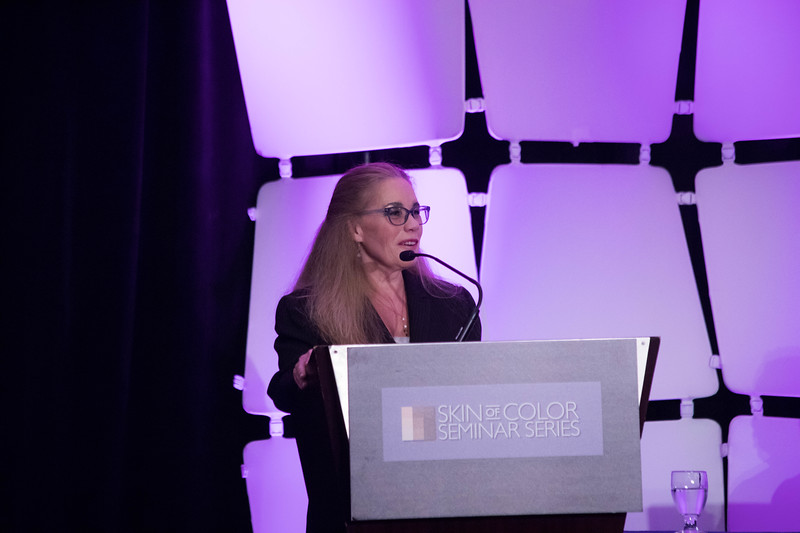
Wendy Roberts Presenting at SOCU

Source: Dermatology News

This is an excerpt from Dermatology News’ coverage of Skin of Color Update 2019.

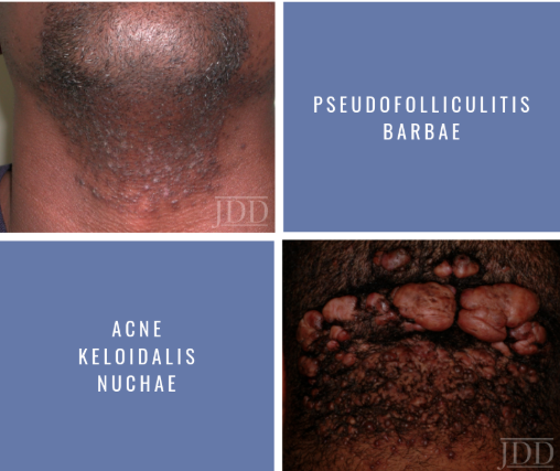
For women with pseudofolliculitis barbae, an empirically-based strategy of microdermabrasion, laser treatment, emollients, and maintenance retinoids has been found highly effective, Wendy Roberts, MD, reported at the Skin of Color Update 2019.

“We didn’t have great treatments for this problem in the past, but the technology has evolved, and you can now get most women clear,” Dr. Roberts, a dermatologist who practices in Rancho Mirage, Calif., said at the meeting.

This approach is appropriate in all women, but Dr. Roberts focused on her experience with black patients, for whom an antioxidant cream is added to address the inflammatory-associated hyperpigmentation that often accompanies pseudofolliculitis barbae, a chronic inflammatory skin condition typically characterized by small, painful papules and pustules.

Start with microdermabrasion to treat the hypertrophic hair follicles and address keratin plugs, Dr. Roberts said. The microdermabrasion smooths the skin and increases penetration of subsequent creams and topics, she said.

“In the same session, I treat with Nd-YAG 1064 nm laser using short pulses,” she noted. For black women, she makes four passes with the laser at a level of moderate intensity. For those with lighter skin, she might perform as many as six passes with the laser set higher.

The microdermabrasion is repeated monthly for three or four treatments, but can be extended for those with persistent symptoms, Dr. Roberts pointed out. She presented a case of a patient who required seven treatments to achieve a satisfactory response.

Read More….

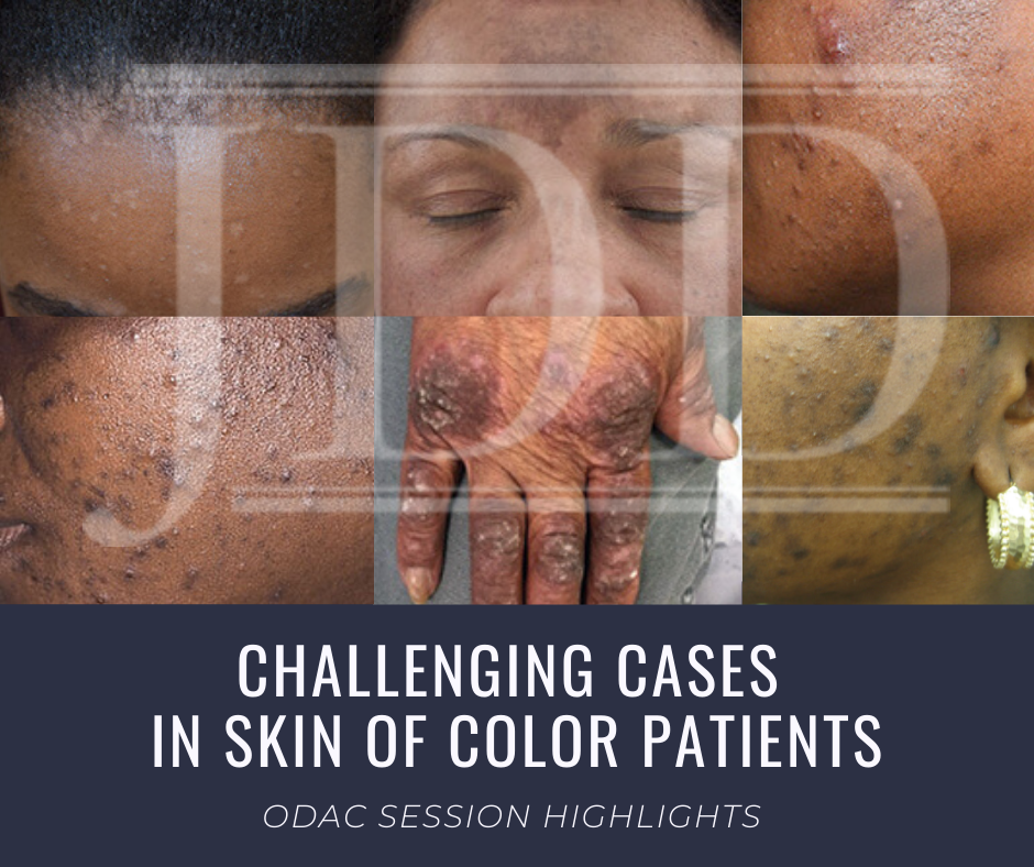
Challenging Cases in Skin of Color Dermatology Patients

By Media Coverage, Medical Dermatology, Sessions
Skin of Color patient dermatology cases

Source: Next Steps in Derm

This year at the 17th Annual ODAC Dermatology, Aesthetic & Surgical Conference (ODAC), Dr Amy McMichael presented the audience with new pearls of advice on how to approach and diagnose complex medical dermatology cases in patients with skin of color. During her session, she addressed the important need for providers to be able to recognize disease in patients of all races. The majority of the global population consists of people with skin of color and the US population is changing to include a higher percentage of patients with diverse backgrounds. She covered a wide range of diagnoses from psoriasis to melasma and how these may present differently is darker skin types. As she walked the audience through each case it became apparent that being able to recognize and treat certain conditions in patients with skin of color is not only essential but also complex in nature.

First, Dr McMichael summarized the top conditions that African American patients were evaluated for during a dermatologist visit. The top 6 conditions included:

This helped to set the scene for the first case involving a 40-year-old African American female with hidradenitis suppurativa presenting with draining gluteal plaques. Even though the biopsy showed granulomatous dermatitis, the patient was not improving with multiple treatments and developed worsening pain and drainage from gluteal plaques. On a second biopsy the pathology showed psoriasis with granulomatous changes. The patient eventually improved with the systemic treatment Humira, a TNF-a inhibitor. Her major takeaways from this case included:

  • Do a second biopsy if the patient’s skin is not responding as expected to the treatment you have prescribed
  • Psoriasis can have a unique presentation similar to existing hidradenitis
  • Use systemic treatments early to help control symptoms

Second, she tackled the challenge of treating melasma with combination therapies. In melasma, there is too much melanin being created by melanocytes and it is then carried by keratinocytes. These cells then release melanin into the dermis, causing blotchy pigmentation often on the face. Topical therapies are usually directed towards preventing increased creation of melanin by melanocytes. People often use hydroquinone 2% or 4% along with encouragement of consistent daily sunscreen use. If used at too high of a concentration, then hydroquinone may cause ochronosis (skin becomes bluish – grey).

Dr McMichael suggested adding a novel treatment called cysteamine to the regimen for melasma treatment for more effective results. Cysteamine is an aminothiol that is made in our cells from the amino acid cysteine. Although more interest is arising now for its use in treating melasma, cysteamine was actually researched in 1966 when scientist Dr Chavin injected it into black goldfish skin and observed partial depigmentation. Cysteamine 5% cream may be a more effect treatment for melasma with less side effects.

Another novel treatment Dr McMichael discussed was the use of tranexamic acid for resistant melasma. This is another derivative of an amino acid, lysine, and it works as an anti-fibrinolytic. It has the ability to block UV-induced plasmin activity within keratinocytes. Patients would need to be screened out by their providers for a past medical history of DVT, pulmonary embolism, heart disease, and stroke before starting the oral medication. She emphasized the importance of getting a good medical history related to these conditions since tranexamic acid could increase the risk of these conditions. For patients who are able to take the medication they are expected to experience a few side effects such as mild GI upset and palpitations. This medication could provide improvement for many patients with chronic melasma who have had to struggle with this condition.

Third, in the next case we were reminded by Dr McMichael that keloids can be very disfiguring and distressful to patients. She talked about using intralesional Kenalog with contact cryotherapy as effective treatments of keloids. Other options for treatment included combining cryosurgery, intralesional Kenalog, and doxycycline. It was eye opening for the audience to hear her say we should be thinking about keloids not just as scars but tumors representing overgrowth of tissue. This paradigm shift of how we think about keloids can further shape how we think about treatment modalities for keloids as well.

Read More….

Dermatology Concerns In Skin of Color Patients

By Skin of Color Update Agenda
Acne Keloidalis Nuchae

During the 16th Annual ODAC Dermatology, Aesthetics and Surgical Conference, I had the pleasure of taking part in the Resident Career Development Mentorship Program (a program supported by an educational grant from Sun Pharmaceutical Industries, Inc.). and was paired with Dr. Andrew Alexis, Chair of Dermatology at Mount Sinai St. Luke’s and Mount Sinai West in New York City.

During a 45-minute working group session, Dr. Alexis covered three main themes: common dermatologic disorders with unique manifestations in skin of color, disorders that disproportionately affect patients of color and therapeutic nuances and unique treatment concerns in skin of color. Here are the main takeaways and pearls from this session.

Common Disorders With Unique Manifestations

Acne

Post-inflammatory hyperpigmentation is a major concern in patients of color and many times more bothersome than acne itself.  It is important to use agents that will treat both acne and PIH. Retinoids can be very effective –tretinoin, tazarotene and adapalene have been shown effect for PIH. Azelaic acid can be a good add on for patients with PIH.

Dr. Alexis doesn’t use much hydroquinone for these patients – the macules left behind by acne are usually too small to avoid creating halos around the lesion.  Chemical peels have been demonstrated to improve PIH in small studies.  The risks are higher in skin of color, so it advisable to stick to superficial peeling agents. Avoiding irritation is essential since it can lead to more dyspigmentation.

Maximize tolerability – adapalene and low concentration tretinoin or tazarotene are a good starting point.  Eliminate any irritating scrubs and other skincare products.  Use noncomedogenic moisturizers concurrently.

Disorders That Disproportionately Affect Patients of Color

Pseudofolliculitis barbae

Findings consist of papules and prominent hyperpigmentation.  This process can also trigger keloid formation. While more common in men, women with hirsutism may also develop PFB. It results from a foreign body reaction to hair reentering the dermis.

A very effective strategy is to discontinue shaving.  You may have to write letters to some patients’ employers in order to excuse them from shaving (Dr. Alexis keeps a form letter on file in his practice).

Chemical depilatory agents are a decent option.  Barium sulfide powder and calcium thioglycolate cream can be used every 2-4 days.  However, they can cause irritant dermatitis. Some patients may also find success by modifying their shaving practices.  Don’t assume your patients know how to shave – educate them.  Electric clippers are a good option – have patients leave 0.5-1 mm stubble. Traditionally single blade manual razors have been recommended.

One study sought to quantify the impact of blade number on PFB – 90 African American men were assigned to shave with a different number of blades.  There was no difference between any of the groups and everyone got better.

A small study showed decreased severity of PFB with daily shaving vs twice weekly shaving.

Dr. Alexis has a handout for patients with shaving instructions: before shaving use a mild cleanser and use a wash cloth in a circular motion to free hairs.  Use clean and sharp razor, shaving in the direction of hair growth.  Use topicals after such as clindamycin lotion or topical dapsone.  Apply a topical retinoid nightly. Avoid pulling or plucking embedded hairs, shaving against the grain.

Read More

Skin of Color Update Co-Chair Dr. Eliot Battle Shares Insights into 2019 Faculty and Topics

By Sessions, Skin of Color Update Agenda

Skin of Color Update Co-Chair, Dr. Eliot Battle, discusses the elite faculty lineup and topics planned this year including hair loss, keloids, rosacea, acne, lasers, aesthetic treatments, skin cancer, medical dermatology, melasma, hyperpigmentation, vitiligo, inflammatory diseases and much, much more!

Skin of Color Update 2019 (previously Skin of Color Seminar Series) is the largest CE event dedicated to trending evidence-based research and new practical pearls for treating skin types III – VI. Attendees leave with critical annual updates and fresh practical pearls in skin of color dermatology.

Join us this year in New York City, September 7-8, 2019! Register today at https://skinofcolorupdate.com/registration-hotel-2019/

Co-Chair Dr. Alexis Shares the Exciting 2019 Program Highlights

By Sessions, Skin of Color Update Agenda

Skin of Color Update 2019 (previously Skin of Color Seminar Series) is the largest CE event dedicated to trending evidence-based research and new practical pearls for treating skin types III – VI. Attendees leave with critical annual updates and fresh practical pearls in skin of color dermatology. Earn CE in New York City with direct access to elite experts and an experience unmatched by any other event in dermatology.

Keloids skin of color update Baldwin

Updates on the Prevention and Management of Earlobe Keloids

By Skin of Color Update Agenda

This article features a recap of Dr. Hilary Baldwin’s talk on the etiology, risk factors, and treatment of keloids at the 2018 Skin of Color Seminar Series, now known as the Skin of Color Update. Dr. Bridget Kaufman, onsite correspondent for the meeting, shares highlights directly from the talk. Dr. Baldwin focused on earlobe keloids (cartilage piercing keloid) in particular, which may present with several different morphologies: anterior button, posterior button, wraparound, dumbbell, and lobular. *Clinical pearls* from this session are bolded, underlined and marked with asterisks.

Some patients develop cartilage keloids and others do not. Based on a study of 220 patients at Kings County Hospital, there appears to be no difference in rate of cartilage piercing, metal sensitivity, types of earrings worn, piercing method, hormonal influences, or age at piercing between keloid formers and non-keloid formers.  In the keloid former group, 12.8% of patients developed keloids at the first piercing, and the risk of keloid formation dramatically increased at each piercing thereafter (70.2% risk for 2nd piercing).

Important Points:

*Ear pierces on babies have a 0% risk of keloids*

*Piercings done pre-menarche have a significantly lower risk of keloids than those performed post-menarche*

*First pierces rarely keloid*

*The chances of subsequent pierce keloiding in a keloid former is at least 20% or higher*

*Earlobe keloids are significantly easier to treat than classic keloids on the body.* Earlobes are a discrete tissue with little to no tension, pressure dressings can be used, and patients tend to be very motivated and compliant. *For surgery alone, recurrence rate on the ear is 39-42% (vs. 100% on body). Surgery plus corticosteroids and surgery with radiation therapy for earlobe keloids are associated with a 1-3% (vs. 50% on body) and 0-25% (same for body) risk of recurrence.* Imiquimod helps prevent keloid recurrence when used on the earlobes, but does not appear to be effective on other body parts.

Dr. Baldwin’s method for excising dumbbell keloids called *dumbbell keloids for dummies:*

  • Shave off anterior button
  • Shave off posterior button
  • Measure diameter of keloid core
  • Select punch to be at least 1mm wider than core
  • Stabilize and punch through to a tongue depressor
  • Suture right/left anteriorly and superior/inferior posteriorly

Unfortunately, not all keloids can be surgically excised. The major dogma of keloid surgery is “don’t leave any keloid tissue behind.” *Hilary’s dogma of earlobe keloid surgery is “a non-functional earlobe is a treatment failure.* Dr. Baldwin performed a study in 5 patients in whom complete removal of keloids would leave a non-functional earlobe. She sculpted the keloid to earlobe shape, injected interferon-alpha 2b 1.5-million units/cm, covered the wound with a compression earring, and allowed for secondary intention healing. In the patients who completed therapy, there was no uncontrollable recurrence at 4-6 year follow-up, although all patients are continuing the use of pressure earrings. Based on these results, *the combination of targeted keloid removal, interferon-alpha 2b injection, and pressure earrings may be an option for patients with large, difficult-to-treat keloids.*

Read more.


Register for Skin of Color Update for lectures and pearls on keloids and more.

Successful Treatment of Keloid With Fractionated Carbon Dioxide (CO2) Laser and Laser-Assisted Drug Delivery of Triamcinolone Acetonide Ointment in an African-American Man

By SOC Manuscripts

Source: J Drugs Dermatol. 2017;16(9):925-927.

Ekaterina Kraeva MD, Derek Ho MD, and Jared Jagdeo MD MS

Keloids are fibrous growths that occur as a result of abnormal response to dermal injury. Keloids are cosmetically disfiguring and may impair function, often resulting in decreased patient quality-of-life. Treatment of keloids remains challenging, and rate of recurrence is high. We present a case of a 39-year-old African-American man (Fitzpatrick VI) with a 10-year history of keloid, who was successfully treated with eight sessions of fractionated carbon dioxide (CO2) laser immediately followed by laser-assisted drug delivery (LADD) of topical triamcinolone acetonide (TAC) ointment and review the medical literature on fractionated CO2 laser treatment of keloids. To the best of our knowledge, this is the first report of successful treatment of a keloid using combination therapy of fractionated CO2 laser and LADD with topical TAC ointment in an African-American man (Fitzpatrick VI) with excellent cosmetic results sustained at 22 months post-treatment. We believe that this combination treatment modality may be safe and efficacious for keloids in skin of color (Fitzpatrick IV-VI) and other patients. This case highlights the ability of laser surgeons to safely use fractionated CO2 lasers in patients of all skin colors.

Read more.