Category

Sessions

Ethnicity Matters: Medical Dermatology Concerns Across Ethnic Groups

By Medical Dermatology, Sessions
Wendy Roberts Presenting at SOCU

Source: Dermatology News

This is an excerpt from Dermatology News’ coverage of Skin of Color Update 2019.

For women with pseudofolliculitis barbae, an empirically-based strategy of microdermabrasion, laser treatment, emollients, and maintenance retinoids has been found highly effective, Wendy Roberts, MD, reported at the Skin of Color Update 2019.

“We didn’t have great treatments for this problem in the past, but the technology has evolved, and you can now get most women clear,” Dr. Roberts, a dermatologist who practices in Rancho Mirage, Calif., said at the meeting.

This approach is appropriate in all women, but Dr. Roberts focused on her experience with black patients, for whom an antioxidant cream is added to address the inflammatory-associated hyperpigmentation that often accompanies pseudofolliculitis barbae, a chronic inflammatory skin condition typically characterized by small, painful papules and pustules.

Start with microdermabrasion to treat the hypertrophic hair follicles and address keratin plugs, Dr. Roberts said. The microdermabrasion smooths the skin and increases penetration of subsequent creams and topics, she said.

“In the same session, I treat with Nd-YAG 1064 nm laser using short pulses,” she noted. For black women, she makes four passes with the laser at a level of moderate intensity. For those with lighter skin, she might perform as many as six passes with the laser set higher.

The microdermabrasion is repeated monthly for three or four treatments, but can be extended for those with persistent symptoms, Dr. Roberts pointed out. She presented a case of a patient who required seven treatments to achieve a satisfactory response.

Read More….

Long-Term Benefits of Daily Photo-Protection With a Broad-Spectrum Sunscreen in United States Hispanic Female Population

By Aesthetic Dermatology, Medical Dermatology
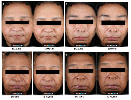
Image of photo aging

Source: JDD Online

The following is an excerpt from the Journal of Drugs in Dermatology article, Long-Term Benefits of Daily Photo-Protection With a Broad-Spectrum Sunscreen in United States Hispanic Female Population.

Introduction
The demographics of the United states are evolving with a large increase in racial and ethnic diversity driven by international migration of Hispanic, African, and Asian populations leading to a minority-majority shift in ~2050 towards persons of color (Fitzpatrick III, IV, V, and VI).1 Specifically, the Hispanic population is projected to be among the fastest growing population in the US, projected to increase from 55 million in 2014 to 119 million in 2060, a change of +115%.1

Subjects with skin of color are heterogeneous with multiple shades and tones and different reactions to intrinsic and extrinsic aging factors due to structural and physiologic differences.2,3 Skin of color individuals have fewer visible signs of aging (deep wrinkles, fine lines, rough surface texture, and sun spots). However, darker skin tones are more susceptible to certain skin conditions including post-inflammatory hyperpigmentation (may occur after acne, eczema, injury, laceration, melasma, post-inflammatory hypopigmentation, pityriasis alba (round, light patches covered with fine scales), dry or “ashy” skin, dermatosis papulosa nigra, and/or greater risk of keloid development.2,3 The incidence of skin cancer among US Hispanics has also increased 1.3% annually from 1992 to 2008.4

Photodamage is characterized histologically by degeneration of the connective tissue and abnormalities in keratinocytes and melanocytes. Clinically, it manifests primarily with wrinkles, dyschromia, texture changes, and, in more severe cases skin cancer.5 Formulations containing broad spectrum sunscreens against both UVA and UVB play an essential role in the prevention of photodamage and UV-induced skin cancers.6,7,8 However, the majority of clinical research on photoprotection has been conducted on subjects with Fitzpatrick types I to III skin and have reported improvements in signs associated with skin aging and texture.9,10 Verschoore et al was the first to conduct a short-term clinical study in India with Phototype IV and VI subjects, and provided first evidence on the effectiveness of daily sunscreen use on skin tone and radiance.11 Similar benefits were observed in an 8-week study in US.12

Although sun protection is highly recommended by dermatologists for skin cancer risk-reduction and the prevention of premature aging or pigmentary disorders, adherence to the recommendations is not commonly observed among US Hispanics.13 Moreover, a large number of US Hispanics reside in areas with high UV index with a high degree of sun seeking behavior. Among Hispanic adults who report engaging in sun protection, they do so mostly by staying in the shade (53.7%) rather than use of sunscreen (32.3%) or wearing sun protective clothes (18.1%); while 36.7% of the subjects surveyed indicated that they never use sunscreen.14,15 There are sociodemographic factors that contribute to the adherence to safe sun behaviour such as education, age, and gender, etc, therefore there is a need to raise awareness of skin cancer risks, advocate for preventive measures and educate on benefits of sunscreen and sun protection among US Hispanics.16

The benefits of topical agents for reversal of sun damage has been well established. Use of retinoic acid and its derivatives or other drugs to reverse and improve sun damaged skin has been demonstrated in many studies.17,18 Long-term sunscreenuse along with other topical agents have also been shown to prevent photodamage and hyperpigmentation in fair-skinned subjects.19 For effective photoprotection, sunscreen products containing both SPF and PPD are essential to battle the harmful UVB (skin cancer risks) and UVA (photo-aging risks).20 Daily use of a broad-spectrum sunscreen (SPF 30) over a one-year period has also been demonstrated to improve clinical parameters of photodamage in phototype I-III subjects.10 However, a comprehensive long-term sunscreen use study in skin of color is lacking. Therefore, this study was designed to assess the benefits of sunscreen of SPF30/PPD 20 in Hispanic women of Fitzpatrick skin types IV and V over 12 months in comparison to a real-life observational group with subjects who did not use sunscreen regularly.

Read Full Article….

Discussions and Conclusions
Effective photoprotection is critical for healthy skin, in preventing skin cancers, reducing photodamage, and improving aesthetic appearance. A broad spectrum sunscreen protecting against both UVA and UVB irradiation is essential. Protecting against the UVA spectrum needs special attention, especially under daily diffused exposure, as UVA is more penetrating and less affected by seasonality and impacts photoaging and skin oxidative stress.22 It has been reported that in order to receive effective photoprotection on skin, a PPD value of 18 is desired.20 In this study, the investigational product with SPF 30/PPD 20 is considered sufficient for daily activity without prolonged direct sun exposure when applied properly. Concerning skin of color population, the use of sunscreen is lower than in Caucasians despite high prevalence of sun-related pigmentary disorders and rising rates of cutaneous cancers.4 This study provides strong evidence to educate and advocate for daily use of a proper sunscreen product for populations with high phototype skin.

The clinical evaluation demonstrated significant visible improvement in sunscreen group starting from 3 months and progressive increased over time. Benefits on multiple facial areas and body sites were visible (upper, mid- and lower face, neck, and hands), not only on pigmentary-related concerns (skin tone evenness, overall hyperpigmentation, dark spots, and blotchiness), but also on aging parameters such as fine lines, skin texture, and overall skin quality. This suggests that beyond the preventative benefits, long-term persistent use of a proper sunscreen may also allow the photodamaged skin to self-heal and repair over time.

Histological observations further supported the clinical findings. The observation that the real-life group had higher tendency for pigmentation incontinence is of strong research interest. It has been reported that UV irradiation can destabilize and damage the dermal-epidermal junction (DEJ), which facilitates the entrapment of melanin in the dermis.23 The dermal melanin is extremely difficult to remove, often resulting in stubborn hyperpigmentation.24 This is especially important for skin of color population in whom dermal hyperpigmentation lesions are common and can be worsened with excessive sun exposure. This study provides the first evidence that effective daily photoprotection can be a strategy to prevent dermal melanin formation by protecting the DEJ. A larger sample size study with DEJ biomarkers will help to further elucidate this hypothesis. Infiltration of CD68-positive Macrophages is a hallmark of the inflammatory response after UV irradiation. In the dermis, 2 out of 3 of the real-life biopsy samples showed significant increase in CD68 positive macrophage cells at 12 months compared to baseline, while such change was not observed in the sunscreen group. This suggests the potential preventative benefits of sunscreen in subclinical skin inflammation induced by chronic exposure to UV. In all of the histological evaluations, thegeographical location in which the study was conducted (Los Angeles versus Washington, DC) was not a strong contributing factor to any of the observed differences. However, the histological findings in this study are limited by the small number of biopsies obtained.

In summary, this 12-month study on long-term persistent use of an SPF30/PPD20 sunscreen on phototype IV and V subjects demonstrated significant improvement in skin quality and improvement in skin color and photoaging parameters. To our knowledge, this is the first study of this kind in skin of color and Hispanic population. This study confirms that effective sunscreen use is not only protective and beneficial for light skin population but is also critical in improving skin condition for skin of color patients. Overall, the study demonstrates that daily use of sunscreen can protect skin from photo related damage and even reverse some of the photo-damage that has already occurred in skin. In addition to previous studies that demonstrated the photo-protective properties of sunscreen use in normal and diseased skin states7,8,9,10 and in view of the fact that good photoprotection behaviors are not common among Hispanics,14,15,16 studies of this type can help educate and stress the importance of daily use of sunscreen and other sun protection behaviors in Hispanic and other skin of color populations.

Read Full Article….

Intralesional Triamcinolone Acetonide in the Treatment of Traction Alopecia

By Aesthetic Dermatology, Case Reports, Medical Dermatology, Sessions
Patient with Alopecia

Source: Next Steps in Derm

In this case series, JDD authors evaluate the efficacy and safety of intralesional triamcinolone acetonide injections (ILK) when used with topical minoxidil in the management of traction alopecia in 6 African American women.

Background

Traction alopecia (TA) is a form of hair loss secondary to repetitive and/or prolonged tension to a hair follicle over an extended period of time. This typically results from wearing tight hairstyles, or an acute traumatic event.1,2 As the etiology is mechanical trauma of the hair follicle, it can occur in any ethnic/racial demographic or gender. It has been observed in ballerinas, as well as Sikh Indian males, all of whom wear hairstyles that exert tension on the frontotemporal hairline. However, most cases of TA occur in women of African descent.1,3

The diagnosis of TA can be made clinically, as well as through the histological examination of a scalp biopsy. The earliest signs of TA are perifollicular erythema and pruritus with or without surrounding papules and pustules.4 The fringe sign of TA is a clinical finding characterized by the presence of retained hair along the frontal and/or temporal hairline, and it has been shown to have high sensitivity for detecting early and late disease of TA.5 On dermoscopy, one may observe reduced hair density with an absence of follicular openings in late stages, and in earlier stages an absence of hairs with preserved follicular openings outlined in brown, particularly at the periphery of the patch of affected scalp, corresponding to the pigmented basal cell layer of the follicular infundibulum that can be seen on histology.6,7 The histological findings can also vary depending on the stage of the disease. Early findings on histology include trichomalacia, normal number of terminal hairs, preserved sebaceous glands, and increased number of telogen and catagen hairs.8 Late disease findings include a decreased number of terminal hair follicles which have been replaced by fibrous tracts, vellus hairs, and retained sebaceous glands.8

Recommended treatment for traction alopecia includes the use of minoxidil and intralesional steroid injections. However, evidence-based proof of the efficacy of ILK in the improvement of TA has not been reported in the literature. In this case series, we evaluate the efficacy and safety of intralesional triamcinolone acetonide injections (ILK) when used with topical minoxidil in the management of TA in 6 African American women.

Methods

A retrospective chart review was performed in patients carrying a diagnosis of TA, who were seen at an active hair disorder clinic between January 2016 and December 2017. All patients who were treated with ILK, and whose treatment progress were recorded with photographs were included. Those who used minoxidil as an adjunct treatment were also noted. The management of TA was assessed by comparing the changes in hair density along the frontotemporal hairline. All patients had been instructed to avoid tension-related hair care practices.

Discussion

This study shows that ILK, when used in conjunction with topical minoxidil, is effective in halting TA progression, and in improving frontotemporal hair density in patients with TA. Our patients reported no adverse systemic effects from the injections that are commonly associated with corticosteroids, and only one patient reported itch in the frontotemporal hairline, a symptom which is more likely a side effect of the topical minoxidil or a manifestation of the TA pathology itself.

Results

Of the TA patients seen, 6 met the criteria for our observational study. All 6 were African American females presenting for evaluation of frontotemporal hair loss, with ages ranging from 32 to 61 years. All subjects reported a history of hairstyling that exerted tension to the frontotemporal hairline at some point in their lives, whether it was recent, during childhood, or both. The clinical diagnosis of TA was established through the presence of the fringe sign. Five subjects had 3 to 4 ILK injections done at 6 to 8-week intervals, performed at a concentration of 5 mg/mL, while one subject (Subject #2) received only one treatment with ILK (Table 1) also at a concentration of 5 mg/mL. Injections were done both at the border of the hair loss in the frontotemporal hairline and extending backwards to include the normal density hair. Subjects concurrently used topical minoxidil 5% daily, and one subject (Subject #2) also took oral doxycycline. All subjects reported the cessation of all hair care practices that exert tension to the frontotemporal hairline, including tight ponytails, tight hair braiding/weaving, twisting of locks, use of scarves to tie hair down, and the use of hair gel on the frontotemporal hairline. All subjects demonstrated a visible increase in hair density along the frontotemporal hairline following their third treatment (Figure 1). None of the subjects reported any serious adverse effects from the injections. The subject that received only one ILK treatment and continued dual therapy on minoxidil and doxycycline reported itch initially, which was improved with the use of a topical steroid.

Read More….

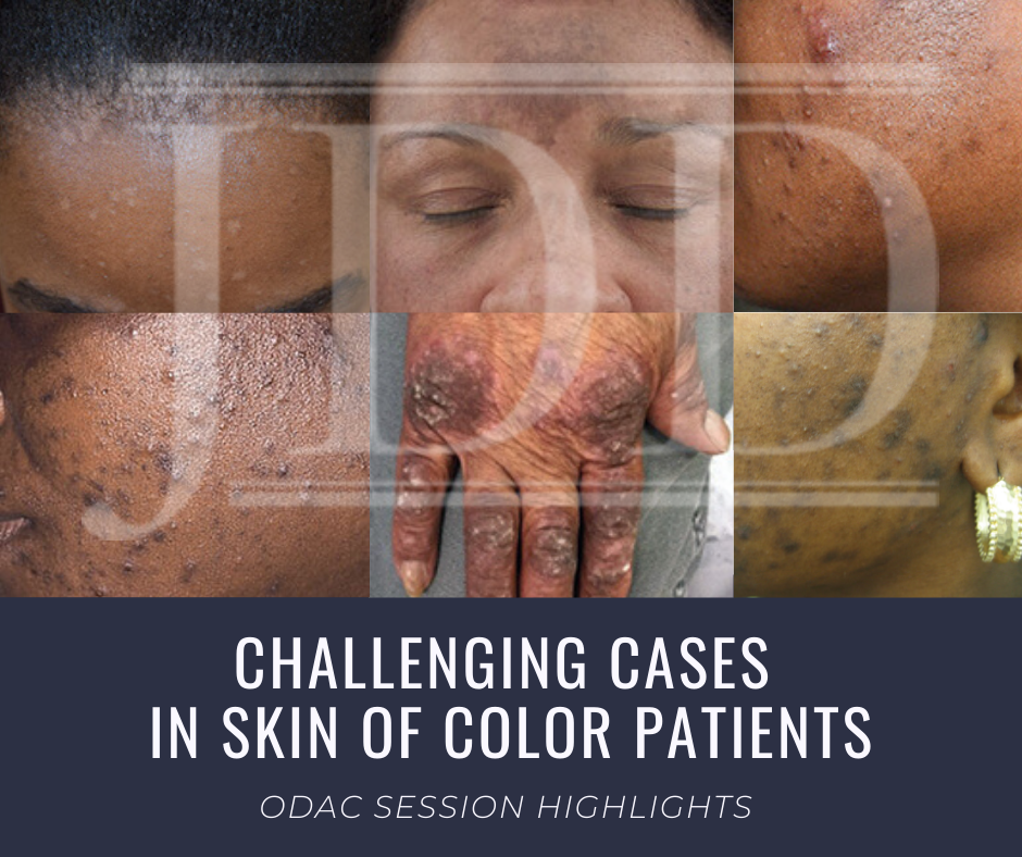
Challenging Cases in Skin of Color Dermatology Patients

By Media Coverage, Medical Dermatology, Sessions
Skin of Color patient dermatology cases

Source: Next Steps in Derm

This year at the 17th Annual ODAC Dermatology, Aesthetic & Surgical Conference (ODAC), Dr Amy McMichael presented the audience with new pearls of advice on how to approach and diagnose complex medical dermatology cases in patients with skin of color. During her session, she addressed the important need for providers to be able to recognize disease in patients of all races. The majority of the global population consists of people with skin of color and the US population is changing to include a higher percentage of patients with diverse backgrounds. She covered a wide range of diagnoses from psoriasis to melasma and how these may present differently is darker skin types. As she walked the audience through each case it became apparent that being able to recognize and treat certain conditions in patients with skin of color is not only essential but also complex in nature.

First, Dr McMichael summarized the top conditions that African American patients were evaluated for during a dermatologist visit. The top 6 conditions included:

This helped to set the scene for the first case involving a 40-year-old African American female with hidradenitis suppurativa presenting with draining gluteal plaques. Even though the biopsy showed granulomatous dermatitis, the patient was not improving with multiple treatments and developed worsening pain and drainage from gluteal plaques. On a second biopsy the pathology showed psoriasis with granulomatous changes. The patient eventually improved with the systemic treatment Humira, a TNF-a inhibitor. Her major takeaways from this case included:

  • Do a second biopsy if the patient’s skin is not responding as expected to the treatment you have prescribed
  • Psoriasis can have a unique presentation similar to existing hidradenitis
  • Use systemic treatments early to help control symptoms

Second, she tackled the challenge of treating melasma with combination therapies. In melasma, there is too much melanin being created by melanocytes and it is then carried by keratinocytes. These cells then release melanin into the dermis, causing blotchy pigmentation often on the face. Topical therapies are usually directed towards preventing increased creation of melanin by melanocytes. People often use hydroquinone 2% or 4% along with encouragement of consistent daily sunscreen use. If used at too high of a concentration, then hydroquinone may cause ochronosis (skin becomes bluish – grey).

Dr McMichael suggested adding a novel treatment called cysteamine to the regimen for melasma treatment for more effective results. Cysteamine is an aminothiol that is made in our cells from the amino acid cysteine. Although more interest is arising now for its use in treating melasma, cysteamine was actually researched in 1966 when scientist Dr Chavin injected it into black goldfish skin and observed partial depigmentation. Cysteamine 5% cream may be a more effect treatment for melasma with less side effects.

Another novel treatment Dr McMichael discussed was the use of tranexamic acid for resistant melasma. This is another derivative of an amino acid, lysine, and it works as an anti-fibrinolytic. It has the ability to block UV-induced plasmin activity within keratinocytes. Patients would need to be screened out by their providers for a past medical history of DVT, pulmonary embolism, heart disease, and stroke before starting the oral medication. She emphasized the importance of getting a good medical history related to these conditions since tranexamic acid could increase the risk of these conditions. For patients who are able to take the medication they are expected to experience a few side effects such as mild GI upset and palpitations. This medication could provide improvement for many patients with chronic melasma who have had to struggle with this condition.

Third, in the next case we were reminded by Dr McMichael that keloids can be very disfiguring and distressful to patients. She talked about using intralesional Kenalog with contact cryotherapy as effective treatments of keloids. Other options for treatment included combining cryosurgery, intralesional Kenalog, and doxycycline. It was eye opening for the audience to hear her say we should be thinking about keloids not just as scars but tumors representing overgrowth of tissue. This paradigm shift of how we think about keloids can further shape how we think about treatment modalities for keloids as well.

Read More….

Rosacea in Skin of Color

Rosacea in Skin of Color

By Medical Dermatology, Sessions

What is Rosacea? 

Rosacea is a common chronic inflammatory skin disease that primarily impacts the face, and includes papules, pustules, erythema, telangiectasias, perilesional redness, phymatous changes, and even eye involvement. Symptoms may vary among different patients and even vary over time in an individual patient. Central facial redness affects many adults and can be an indicator of the chronic inflammatory disease rosacea. Rosacea is a clinical diagnosis based on the patient’s history, physical examination, and exclusion of other disorders.

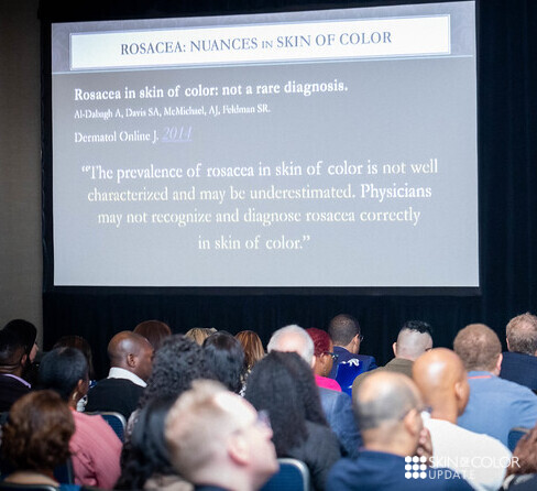
Rosacea is often under-diagnosed, particularly in individuals with skin of color. As a result, Skin of Color Update held a lecture on the topic at the most recent event.

Rosacea: Nuances in Clinical Presentation and Treatment 

At Skin of Color Update 2019, Dr. Fran Cook-Bolden aimed to catch us all up to speed in how to recognize rosacea in more richly pigmented skin. Her lecture on Rosacea: Nuances in Clinical Presentation and Treatment was brimming with practical tips on how to identify the often subtle and overlooked ways that rosacea can manifest in skin of color.  The following is an excerpt of an article by Kimberly Huerth, MD published on Next Steps in Derm.

Because rosacea can have a nuanced presentation in skin of color (SOC), with erythema and telangiectasias that may be difficult to discern in the setting of increased background pigmentation, it was incorrectly assumed for a long time to simply not be there. The reality is that the prevalence of rosacea in SOC is not well characterized but is likely underestimated.1Dermatologists who see a large number of SOC patients, however, will tell you that rosacea is by no means rare in this population. And I am one of these dermatologists—at my Howard University clinic, where I see predominantly black and Hispanic patients, I see several of cases of rosacea every week.

And because a diagnosis not considered is a diagnosis not made, there is often unnecessary progression of rosacea in SOC patients that results from delayed and/or inaccurate diagnosis, which in turn engenders inappropriate or inadequate treatment. As a consequence, this can lead to morbidity in the form of disfiguring, occasionally irreversible cutaneous findings, as well as intense and chronic emotional distress.

Pearls

  • Look for the nuanced clinical findings!!
  • Fixed centrofacial erythema may appear more reddish/violet
  • A patient complains of “acne,” but has no comedones. Additionally, papules and pustules are superimposed on an erythematous background. Inflammatory papules may also appear on the chest and back.
  • Telangiectasias can be difficult to appreciate with the naked eye in FST V – VI, so use your dermatoscope to help you find them
  • Check for scleral injection, which may be a sign of ocular rosacea. Be aware that the onset of ocular findings may proceed cutaneous ones.
  • Phymatous rosacea is a giveaway!
  • Facial edema of the upper 2/3 of the face in a patient who has complained of longstanding rosacea symptoms may represent progression to Morbihan disease. Case in point (quite literally)—a poster that I presented at the Skin of Color Update highlighted a case of Morbihan disease in a black man who had reported symptoms of rosacea to his non-dermatology providers for 16 YEARS before he came to see me and received a correct diagnosis. To learn more about this case, and Morbihan disease, check out Dr. Lola Adekunle’s interview on Next Steps in Derm.
  • Pertinent negatives are just as important as pertinent positives. Know that post-inflammatory hyperpigmentation (PIH) is ALMOST NEVER directly related to rosacea, unless the disease is very chronic and severe OR if the patient has been injured their skin in some way while trying to self-treat disease manifestations (picking at lesions, using harsh topical therapies)

For more rosacea pearls and AHA moments, visit the article on Next Steps in Derm.

Purchase lectures like this one:

Skin of Color Update On Demand Video

Skin of Color in Psoriasis Pearls from SOC Update

Psoriasis in Skin of Color: Pearls from SOC Update

By Sessions, Skin of Color Update Agenda

Andrew Alexis, MD, MPH, co-chair and co-founder of Skin of Color Update presented on Psoriasis in Skin of Color this past event.

Previously thought to be rare in patients of African ancestry, the prevalence of psoriasis is 1.6% in African Americans and 1.4% in Hispanics.

The talk, “Psoriasis: Distinct Clinical Features and Treatment Options of Psoriasis Patients of Color, ” was one of the top rated lectures of Skin of Color Update.

The lecture focused on the fundamentals of dermatology with an emphasis on several key characteristics.

  1. Color and distribution in the clinical presentation
  2. Recognizing common medical mimickers of psoriasis such as lichen planus, sarcoidosis, and cutaneous lupus erythematosus (.e. discoid lupus) in skin of color
  3. When in doubt, do not hesitate to biopsy

During the lecture, Dr. Alexis presented a game of “Psoriasis or Not?” allowing the audience to guess if the Kodachrome was psoriasis.  This illustrated the vast presentation of psoriasis and how papulosquamous disorders can be challenging to differentiate in skin of color.

To read more about this lecture and psoriasis in skin of color, visit the full article on Next Steps in Derm.

Purchase lectures like this one:

Skin of Color Update On Demand Video


Acne in Skin of Color Alexis

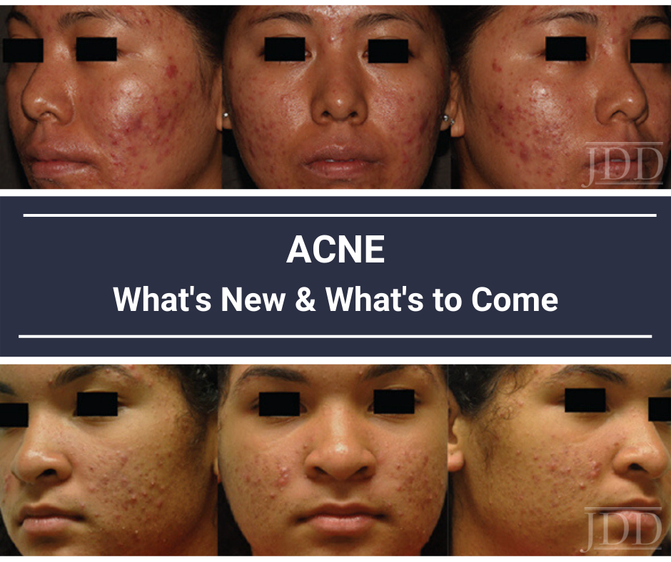
Acne in Skin of Color: What’s New and What’s to Come

By Sessions, Skin of Color Update Agenda

At Skin of Color Update 2019, our co-chair and co-founder, Andrew Alexis, MD, MPH  gave a lecture on Acne: What’s New and What’s to Come?  Our onsite correspondent, Kimberly Huerth, MD, M. Ed, provided the following recap of this is insightful session.  The read the full article, please visit Next Steps in Derm website.   Missed Skin of Color Update 2019? Purchase lectures like this on-demand

By: Kimberly Huerth, MD, M. Ed

I still treat my acne twice daily with a whole cabinet full of various topicals. I’ve tried and failed doxycycline because it disrupts every single molecule of bacterial flora in my body. I’ve tried and failed spironolactone because I was the poster child for nearly all of its annoying and inconvenient side effects. I’ve tried and failed several OCPs because my body was a little too convinced by the estrogen and progesterone that it was actually pregnant, and decided to make me persistently sleepy and nauseous. I could put the 650-microsecond Nd:YAG that we have in clinic to use, but can’t bring myself to bother my co-rezzys (or myself) at the end of a long day seeing patients. And yes, I’ve already done a course of isotretinoin…two courses in fact. And no, I don’t have PCOS. So when I settled in to hear Dr. Andrew Alexis’s lecture on Acne in Skin of Color: What’s new and what’s to come?, I was excited for some new strategies with which to help my patients, and myself.

Dr. Alexis not only shared expert insights and strategies on how to optimize treatment for acne in skin of color (SOC) patients, but he also laid out an overview of some of the new and emerging acne treatments that we will presently be able to add to our armamentarium!

This article will provide an overview of the following:

  • Understanding the unique presentation and needs of SOC patients with acne
  • Sarecycline, a new tetracycline class antibiotic
  • New topical acne medications in the pipeline

But first, let me share a few of the “A-ha” moments that I experienced during Dr. Alexis’s lecture, in the hope that they will entice you to read on…

Read more.


Missed Skin of Color Update 2019? Purchase lectures like this on-demand

Skin of Color Update On Demand Video

Skin of Color Update On Demand Video

SOC Update Launches On-Demand Package

By Sessions, Skin of Color Update Agenda

New York (Nov. 5, 2019) Skin of Color Update launches on-demand video package. Skin of Color Update, the largest CE event dedicated to trending evidence-based research and new practical pearls for treating skin types III – VI expands educational offerings to include on-demand videos.

The demand for skin of color dermatology education is greater than Skin of Color Update’s ability to accommodate onsite. Therefore, SanovaWorks has produced and launched a Best of Skin of Color Update video package.  The video content is comprised of the highest attendee rated sessions of Skin of Color Update 2019.  On-demand videos include top-rated faculty lectures in-sync with PPT slides with accompanying audio files.

On-demand Lectures include:

  • Pearls for the Diagnosis and Treatment of Atopic Dermatitis and Eczema with Andrew Alexis, MD, MPH
  • Complex Medical Cases with Andrew Alexis, MD, MPH and Ted Rosen, MD
  • Surgical Approaches for Keloids with Maritza Perez, MD
  • Hair & Scalp Disorders Treatment Strategies: What, How and When? with Heather Woolery-Lloyd, MD
  • Pearls and Strategies for Preventing Laser Complications with Eliot F. Battle, Jr., MD.

The video content focuses on expert techniques, real-life clinical cases and expert pearls immediately useful in the practice.  For a video preview, click here.

Purchase the on-demand Best of Skin of Color Update video package and start learning from the experts today.

Skin of Color Update On Demand Video

Aesthetic Skin of Color

Consensus and Misconceptions Regarding the Aesthetic Skin of Color Consumer

By Media Coverage, Sessions, Skin of Color Update Agenda

Each month the Journal of Drugs in Dermatology JDD Podcast discusses a current issue in dermatology. During the month of September, podcast host Dr. Adam Friedman sat down with Skin of Color Update 2019 co-chair and co-founder,  Dr. Andrew Alexis, Chair of Dermatology at Mt. Sinai St. Lukes and Mount Sinai West to discuss misconceptions regarding the aesthetic skin of color consumer.

Dr. Angela Hou, PGY-3 dermatology resident at George Washington University School of Medicine and Health Sciences, shares key takeaways from Dr. Alexis’ JDD podcast titled ‘Capturing Consensus and Cutting Out misconceptions regarding the Aesthetic Skin of Color Consumer’.

Here is an excerpt recently published on our media partner, Next Steps in Dermatology.

Key Takeaways

  • There has been a rapid increase in the past 10 years in Fitzpatrick Skin Type IV-VI patient’s seeking aesthetic skin care, however the guidelines for skin of color is limited and more clarification and guidance is needed
  • This article helps reduce the gap in knowledge in regard to skin of color. This was difficult given the lack of evidence-based studies, therefore expert consensus was necessary for deciding on recommendations.
  • A common myth is that darker-skinned patients of African descent do not seek or need injectable fillers of the lips. Although lip enhancement is less common than in other populations, restoration of lip volume is still an important aesthetic concern, albeit at an older age than among Caucasian patients
  • Another knowledge gap is regarding skin of color patients with a history of keloids and the risks of developing keloids after filler injections. However, per the expert consensus, there are no known cases of keloids induced by soft tissue filler injections. Therefore, keloids should not be an absolute contraindication to fillers and should be evaluated on a case-by-case basis.
  • To read more of the key takeaways and words from the investigator, read the full article here.

To hear the JDD podcast, click here.

Purchase

Skin of Color Update On Demand Video