Tag

medical dermatology

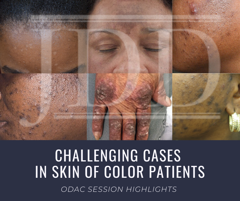
Challenging Cases in Skin of Color Dermatology Patients

By Media Coverage, Medical Dermatology, Sessions
Skin of Color patient dermatology cases

Source: Next Steps in Derm

This year at the 17th Annual ODAC Dermatology, Aesthetic & Surgical Conference (ODAC), Dr Amy McMichael presented the audience with new pearls of advice on how to approach and diagnose complex medical dermatology cases in patients with skin of color. During her session, she addressed the important need for providers to be able to recognize disease in patients of all races. The majority of the global population consists of people with skin of color and the US population is changing to include a higher percentage of patients with diverse backgrounds. She covered a wide range of diagnoses from psoriasis to melasma and how these may present differently is darker skin types. As she walked the audience through each case it became apparent that being able to recognize and treat certain conditions in patients with skin of color is not only essential but also complex in nature.

First, Dr McMichael summarized the top conditions that African American patients were evaluated for during a dermatologist visit. The top 6 conditions included:

This helped to set the scene for the first case involving a 40-year-old African American female with hidradenitis suppurativa presenting with draining gluteal plaques. Even though the biopsy showed granulomatous dermatitis, the patient was not improving with multiple treatments and developed worsening pain and drainage from gluteal plaques. On a second biopsy the pathology showed psoriasis with granulomatous changes. The patient eventually improved with the systemic treatment Humira, a TNF-a inhibitor. Her major takeaways from this case included:

  • Do a second biopsy if the patient’s skin is not responding as expected to the treatment you have prescribed
  • Psoriasis can have a unique presentation similar to existing hidradenitis
  • Use systemic treatments early to help control symptoms

Second, she tackled the challenge of treating melasma with combination therapies. In melasma, there is too much melanin being created by melanocytes and it is then carried by keratinocytes. These cells then release melanin into the dermis, causing blotchy pigmentation often on the face. Topical therapies are usually directed towards preventing increased creation of melanin by melanocytes. People often use hydroquinone 2% or 4% along with encouragement of consistent daily sunscreen use. If used at too high of a concentration, then hydroquinone may cause ochronosis (skin becomes bluish – grey).

Dr McMichael suggested adding a novel treatment called cysteamine to the regimen for melasma treatment for more effective results. Cysteamine is an aminothiol that is made in our cells from the amino acid cysteine. Although more interest is arising now for its use in treating melasma, cysteamine was actually researched in 1966 when scientist Dr Chavin injected it into black goldfish skin and observed partial depigmentation. Cysteamine 5% cream may be a more effect treatment for melasma with less side effects.

Another novel treatment Dr McMichael discussed was the use of tranexamic acid for resistant melasma. This is another derivative of an amino acid, lysine, and it works as an anti-fibrinolytic. It has the ability to block UV-induced plasmin activity within keratinocytes. Patients would need to be screened out by their providers for a past medical history of DVT, pulmonary embolism, heart disease, and stroke before starting the oral medication. She emphasized the importance of getting a good medical history related to these conditions since tranexamic acid could increase the risk of these conditions. For patients who are able to take the medication they are expected to experience a few side effects such as mild GI upset and palpitations. This medication could provide improvement for many patients with chronic melasma who have had to struggle with this condition.

Third, in the next case we were reminded by Dr McMichael that keloids can be very disfiguring and distressful to patients. She talked about using intralesional Kenalog with contact cryotherapy as effective treatments of keloids. Other options for treatment included combining cryosurgery, intralesional Kenalog, and doxycycline. It was eye opening for the audience to hear her say we should be thinking about keloids not just as scars but tumors representing overgrowth of tissue. This paradigm shift of how we think about keloids can further shape how we think about treatment modalities for keloids as well.

Read More….

Lymphomatoid Papulosis JDD Skin of Color

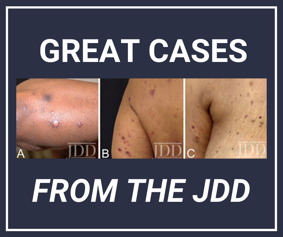
Black Patients with Lymphomatoid Papulosis

By Case Reports, SOC Manuscripts

Lymphomatoid papulosis (LyP) is a CD30+ T-cell lymphoproliferative disorder (LPD) presenting as a recurrent eruption of papules and nodules which resolve spontaneously. CD30+ LPD prevalence in African American (AA)/Black patients is lower compared to White patients. CD30+ LPD has been recently reported to have worse outcomes in AA patients compared to White patients.

A retrospective chart review identified eight AA patients with LyP. Authors Shamir Geller MD, Sarah J. Noor MD, and Patricia L. Myskowski MD describe their experience with these eight patients and review the literature on similar cases.

To view a synopsis of the case published in the Journal of Drugs in Dermatology, visit Next Steps in Derm. Log in to JDDOnline.com for full access of the manuscript.

Racial Differences in Incidence

Major racial differences in incidence among cutaneous lymphoma subtypes have been reported. AA/Blacks have statistically higher incidence ratios of CTCL and MF than other races and a trend towards lower incidence of CD30+ LPD was found in a national US database, which included 31 AAs with CD30+ LPD. A more recent study of another database included 153 AA patients with CD30+ LPD who had a significantly shorter overall survival compared to Caucasians after adjusting for patient disease characteristics, socioeconomic factors and types of treatment.

Discussion

The case series and three additional case reports suggest an indolent disease course of LyP in AA/Black patients. There are several possible explanations for the previous findings on poor survival in AA patients with CD30+ LPD.

  1. These results might be due to inclusion of ALCL and borderline cases with poorer prognosis compared to LyP.
  2. Another possibility is that patients with more aggressive CTCL variants (eg, transformed tumor-stage MF) might have been misdiagnosed or miscoded as CD30+ LPD.
  3. Results support previous reports on an earlier-onset of disease seen in AA/Black patients with LyP5 as well as with other CTCL subtypes, such as MF.  The self-healing crops of papules and nodules can be easily misdiagnosed as other malignant or inflammatory skin conditions (eg, arthropod bites).
  4. The diagnosis of early-stage patch stage MF may be more difficult in Black skin where erythema is less pronounced compared to lighter skin types.

Treatment Approaches

The case series highlights the need for additional studies before clinical recommendation can be made regarding prognosis and treatment in different race groups. Careful physical examination should be performed in Black patients who are diagnosed with LyP and no known history of MF. Once the diagnosis of LyP is made, several treatment approaches are possible.

  1. Noninterventional (“wait-and-see”) strategy is a legitimate approach, especially in patients with a limited number of lesions.
  2. Topical and skin-directed therapies (including topical steroids and phototherapy), and low-dose methotrexate are the best documented therapies for LyP.
  3. There is currently no curative therapy for LyP though the efficacy and safety of brentuximab vedotin, an antibody- drug conjugate directed against CD30, has recently been assessed for the treatment of LyP in 12 patients, including 2 AAs. Brentuximab vedotin was reported to be effective in treating LyP and has been suggested as a possible therapy in severe and refractory cases. Further studies are ongoing to optimize its dosing and to minimize adverse events.

In conclusion, a diagnosis of LyP should be considered in Black patients who present with recurring eruption of papules or nodules that resolve spontaneously. Patients with LyP should be carefully examined for concurrent or later development of MF. Although an indolent course may be expected in Black patients, residual hyperpigmentation and scars following resolution of the LyP lesions are common in this population, highlighting the need for better treatments of this disorder in the Black population.

The Journal of Drugs in Dermatology is available complimentary to US dermatologists, US dermatology residents and US dermatology NP/PA. Create an account on JDDonline.com and access over 15 years of PubMed/MEDLINE archived content.

Patient Buzz: At-Home Laser Hair Removal – The Expert Weighs In

By Media Coverage, Uncategorized
At Home Laser Hair Removal Devices. Are they safe and effective?

Marie Clairerecently posted a list of the magazine’s top devices for at-home laser hair removal, noting their budget-friendly appeal. But are these devices safe and effective? How should you counsel your patients?

For an expert opinion, I consulted dermatologist Eliot F. Battle Jr., MD, CEO and co-founder of Cultura Dermatology & Laser Center in Washington, D.C., clinical instructor in the Howard University Department of Dermatology, and Co-Chair of the Skin of Color Update.

How do at-home laser hair removal devices compare in effectiveness with in-office laser hair removal?

At-home laser hair removal devices have now been available for more than a decade. Just like most gadgets, you get what you pay for, so buyer beware. The devices range from using an intense pulsed light source to using actual diode lasers, although with a much lower energy source then office-based devices. Regardless of which device patients choose, at-home devices do not compare with the efficacy and speed of office-based laser systems. At-home devices are very slow. Because of the amount of time it takes to treat an area and their decrease in efficacy as compared with office-based lasers, I view at-home devices more as “hair-growth delay” devices than “hair-reduction” devices. They can be used alone or as maintenance treatments to office-based hair removal. The main limitations are they are best utilized on smaller areas and are contraindicated on patients with skin of color or tanned skin.

Read More

Skin of Color Update Co-Chair Dr. Eliot Battle Shares Insights into 2019 Faculty and Topics

By Sessions, Skin of Color Update Agenda

Skin of Color Update Co-Chair, Dr. Eliot Battle, discusses the elite faculty lineup and topics planned this year including hair loss, keloids, rosacea, acne, lasers, aesthetic treatments, skin cancer, medical dermatology, melasma, hyperpigmentation, vitiligo, inflammatory diseases and much, much more!

Skin of Color Update 2019 (previously Skin of Color Seminar Series) is the largest CE event dedicated to trending evidence-based research and new practical pearls for treating skin types III – VI. Attendees leave with critical annual updates and fresh practical pearls in skin of color dermatology.

Join us this year in New York City, September 7-8, 2019! Register today at https://skinofcolorupdate.com/registration-hotel-2019/